Trans. Nonferrous Met. Soc. China 23(2013) 996-1001
In vitro corrosion and biocompatibility of phosphating modified WE43 magnesium alloy
Cheng-hong YE1, Ting-fei XI1,2, Yu-feng ZHENG1,3, Shu-qin WANG2, Yang-de LI4
1. Center for Biomedical Materials and Tissue Engineering, Academy for Advanced Interdisciplinary Studies, Peking University, Beijing 100871, China;
2. School of Information and Engineering, Wenzhou Medical College, Wenzhou 325035, China;
3. Department of Advanced Materials and Nanotechnology, College of Engineering, Peking University, Beijing 100871, China;
4. Dongguan EONTEC Co., Ltd., Dongguan 523000, China
Received 10 October 2011; accepted 12 February 2012
Abstract: Phospahting coated WE43 magnesium alloy was prepared by an immersion method. The microstructure, corrosion resistance and biocompatibility of the coated alloy were investigated. Scanning electron microscopy (SEM) and X-ray diffraction (XRD) were used to examine the microstructure and the composition of the coated alloy. The corrosion resistance was studied by means of potentiodynamic polarization method and the biocompatibility of the surface modified WE43 alloy was evaluated by (3-(4,5)-Dimethylthiazol-2, yl)-2,5-diphenyltetrazolium bromide (MTT) and hemolysis test. The results show that the phosphating coating can enhance the corrosion resistance of WE43 alloy and can be a good candidate to increase the biocompatibility of WE43 alloy.
Key words: magnesium alloy; phosphating coating; corrosion resistance; biocompatibility
1 Introduction
Magnesium alloys exhibit an attractive property due to their outstanding biological performance and biodegradability in physiological environment, which makes them become a hot topic in biomedical materials area, especially for implant applications, such as bone [1-4] and intravascular stent [5-8].
However, the low corrosion resistance of magnesium alloys in chloride containing solutions including human body fluid and blood plasma has limited their clinical applications. To improve the corrosion resistance of magnesium alloys, various surface modification techniques, such as alkaline treatment [9], heat treatment [10], micro-arc oxidation [11] and electrodeposition [12], have been studied recently. Among the modification methods especially the chemical treatment method, phosphating may be a promising way for magnesium alloys to improve the corrosion resistance, which remains rarely reported. LIAN et al [13] found that the zinc phosphate coated magnesium alloy exhibited a lower corrosion rate than the untreated substrate, which suggests a higher corrosion resistance of the phosphate coating. ZHANG et al [14] studied the surface characterization of calcium phosphate coated magnesium alloy and found that a Ca�CP coating could provide the magnesium alloy a significant better surface biocompatibility in vitro. In vivo studies also confirmed that the Ca�CP coating exhibited improved osteoconductivity and osteogenesis in the early first four weeks of post-operation period. But in these phosphating studies, a nitrite was used as the accelerating agent which would accelerate the phosphating reaction. However, nitrite is a carcinogen which has been abandoned to use especially in the biomedical application.
In this study, phosphating was used to modify the WE43 magnesium alloy, and sodium m-nitrobenzene sulfonate was used as an accelerator agent of phosphatization to avoid the carcinogen. In addition, previous study has shown that the phytic acid treated magnesium alloy has a better corrosion resistance compared with the untreated alloy [15], which was used in the present phosphating solution. The chemical nature of conversion coating was investigated by both Fourier transform infrared spectroscopy (FTIR) and ESEM equipped with the energy dispersive spectrum (EDS). The corrosion resistance was examined by potentiodynamic polarization method and immersion test in the simulated body fluid (SBF) buffer. The biocompatibility of the modified WE43 magnesium alloy was evaluated by MTT and the hemolysis test, respectively.
2 Experimental
2.1 Preparation of specimens
The WE43 magnesium alloy (91.35% Mg, 4.16% Y, 3.80% RE, 0.36% Zr, 0.20% Zn, 0.13% Mn in mass fraction), provided by Changchun Zhong-Ke-Xi-Mei Magnesium Alloy Co. Ltd., China, was cut into specimens with dimensions of 8 mm in diameter and 1.5 mm in height. The specimens were polished successively with 800#, 1200# and 2000# grit SiC papers, pretreated with acetone and deionized water, dried with hot air, and then immersed in the phosphating solution with compositions of sodium dihydrogen phosphate (NaH2PO4, 20 g/L), phosphoric acid (H3PO4, 6.4 mL/L), sodium metanitrobenzene sulphonate (C6H4NNaO5S, 3 g/L), sodium nitrate (NaNO3, 1.84 g/L), sodium chloride (NaCl, 1 g/L) and phytic acid (2 mL/L). All the reagents were analytical pure (��99%). The conversion coatings were proceeded by immerging the substrate in the solutions with pH 4.5 adjusted by ammonia water, and the temperature stayed at 40 ��C. After immersion, the specimens were thoroughly washed with deionized water and dried overnight at 37 ��C. The untreated specimen was taken as a control.
2.2 Microstructural analysis
The microstructure of the specimens was characterized using an environmental scanning electron microscope (ESEM, AMRAY-1910FE), equipped with the energy dispersive spectrum (EDS). X-ray diffractometer (XRD, Rigaku DMAX 2400) using a Cu K�� radiation was employed to characterize the composition of the coating.
2.3 Immersion test
The immersion test was examined by hydrogen evolution method according to LIU et al [16]. In short, the specimens were put into the center of the beakers filled with the SBF solution, and the hydrogen formed in the corrosion process of WE43 alloy was collected in the funnel which was vertically laid on the surface of the specimen. The corrosion resistance of specimens could be evaluated based on the rate of hydrogen evolution.
2.4 Electrochemical test
The electrochemical measurement was carried out in the SBF solution and kept at (37��0.5) ��C using a water bath. The potentiodynamic polarization curves of the surface coated magnesium alloy were obtained on a three-electrode system. The working electrode was the WE43 magnesium alloy sample with an exposed area of 1 cm2.
2.5 Biocompatibility evaluation
The biocompatibility of the magnesium alloy with or without phosphating modified was evaluated by both cytotoxicity and blood hemolysis experiments. L-929 cells were used to evaluate the cytotoxicity of the experimental samples by the indirect assay. The extract was prepared using Dulbecco��s Modified Eagle��s Medium (DMEM) serum free medium with the surface area of extraction medium ratio 1.5 mL/cm2 in a humidified atmosphere with 5% CO2 at 37 ��C for 72 h. The control groups involved the use of DMEM medium as the negative control and 0.64% phenol DMEM medium as the positive control. L-929 cells were incubated in 96-well culture plates at a concentration of 1��104 mL-1 for 24 h to allow the attachment and the medium was then replaced with 100 ��L extraction. After 1, 2 and 4 d culture, 10 ��L MTT was added to each well cultured for 4 h and 100 mL of the supernatant was measured spectrophotometrically at 570 nm by Elx800 (BioTek instruments). The background with MTT results of extracts was subtracted. In order to evaluate the blood compatibility, samples were soaked in 10 mL normal physiological saline in a tube and kept at 37 ��C for 30 min. 8 mL arterial blood of the volunteer was diluted with 10 mL normal physiological saline that contained 0.5 mL potassium oxalate (20 g/L) decoagulant. After 0.2 mL diluted blood was added into the tube and kept at 37 ��C for 60 min, the tube was centrifugated at 2500 r/min for 5 min. At last, the optical density (D) was obtained with a spectrophotometer (722, Shanghai Precision & Scientific Instrument Co., Ltd, Shanghai, China) at the wave length of 545 nm. The negative and positive groups were 10 mL normal physiological saline with 0.2 mL diluted blood and 10 mL distilled water with 0.2 mL diluted blood, respectively. The hemolysis ratio (R) was calculated according to the following equation: R=[(Dt-Dn)/(Dp-Dn)]��100%, where Dt is the D value of the tested sample, and Dn and Dp are the D values of negative and positive groups, respectively.
3 Results and discussion
3.1 Microstructures
Figure 1 shows the morphology and element composition of the phosphate coating on WE43 magnesium alloy. It can be seen that the coating presents bulk feature and there are few microcracks on the surface of the coating. The chemical composition of the film was analyzed by EDS. From the element compositions we can see that the coating is mainly composed of Mg, F, P, O and C elements, and the presence of P shows that phosphating coating has been successfully coated on the Mg alloy surface, and the F element can be detected too. CUI et al [17], GAO et al [18] and LIANG et al [19] found that magnesium alloy can be modified with phytic acid to form a thin film to slow down the corrosion rate of magnesium alloy, and the P element may suggest that the phytic acid was coated on the WE43 Mg alloy surface.
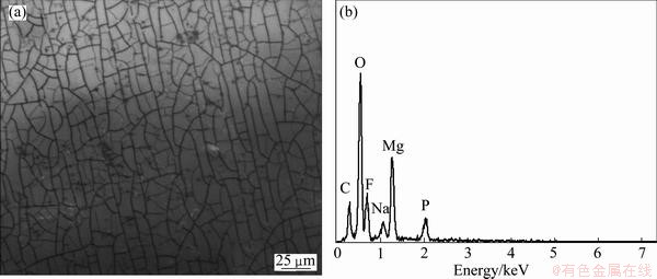
Fig. 1 SEM image (a) and corresponding EDS (b) of phosphating coated WE43 alloy
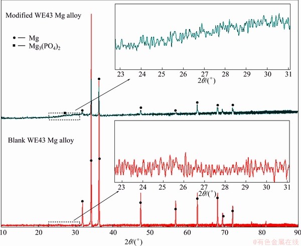
Fig. 2 XRD patterns of WE43 magnesium alloy with and without phosphating
Figure 2 presents the XRD patterns of the phosphating coating on WE43 magnesium alloy. The surface of the blank alloy shows mainly peaks corresponding to magnesium. After exposing to the phosphating media, the intensity of the peaks corresponding to magnesium is lower, indicating the coating on the surface of the alloy, and there is no significant difference between the blank magnesium alloy and the coated magnesium alloy from the XRD patterns. But at 23��-30��, there is an elevating peak which corresponds to the Mg3(PO4)2 phase, compared with the uncoated WE43 alloy.
When the WE43 magnesium alloy was immersed in the phosphating solution, magnesium alloy would dissolve in the solution by the following reaction:
Mg+2H+=Mg2++H2�� (1)
At this pH condition,  should be the major ions, so the Mg2+ dissolving from the magnesium alloy would precipitate on the surface of the alloy as follows:
3Mg2++2��Mg3(PO4)2+4H+ (2)
In the phosphating solution, the accelerator agent nitrate can react with H+ through the following reduction reaction:
 +4H++4e��N2��+O2+2H2O (3)
So, reaction (3) would consume H+ quickly, the local pH at the metal�Csolution interface could be increased quickly, which would facilitate the precipitation of insoluble phosphate [20,21].
In addition, as phytic acid has excellent chelate ability like EDTA, the solution may contain the following reaction:
iMgn++HjPhy(12-j)-=MgiHj(12-2i-j)-Phy (4)
Mg2++2F-��MgF2 (5)
Mg2++F-+ ��MgFPO4 (6)
where Phy represents phytic acid ions, i and j are the different reaction contents [22]. But there is no significant difference between the two XRD patterns, though there exists Mg3(PO4)2. This can be explained by the fact that as the MgiHj(12-2i-j)- Phy complex was chelatoffing the phytic acid and magnesium ions, the complex might be an amorphous structure, which would obscure the other crystal substance, e.g., Mg3(PO4)2, MgF and MgFPO4. Therefore, F element can be found from the EDS image (Fig. 1), but it is not obvious in the XRD patterns.
3.2 Immersion test
Figure 3 shows the hydrogen evolution rate of the uncoated WE43 magnesium alloy and the phosphating coated WE43 alloy in the SBF solution for about 200 h. The overall corrosion reaction of magnesium at its free corrosion potential can be expressed as
Mg+2H2O=Mg2++2OH-+H2�� (7)
1 mol hydrogen evolution would correspond to 1 mol WE43 magnesium alloy during the corrosion, and this result can be a good indicator to evaluate the corrosiveness of magnesium alloys.
The result shows that the coated WE43 magnesium alloy has a slower hydrogen evolution rate while the rate of the blank group is so fast within the beginning of 50 h. Though the two groups have similar evolution rate in the next 160 h, the total volume of the hydrogen evolution of the blank WE43 alloy is more than that of the coated alloy, which suggests a better corrosion resistance of the coated magnesium alloy. This should be because the phosphating coating could well prevent the corrosion reaction, and the coated alloy keeps a stable hydrogen evolution rate, while the uncoated alloy group has a fast corrosion rate in the beginning.
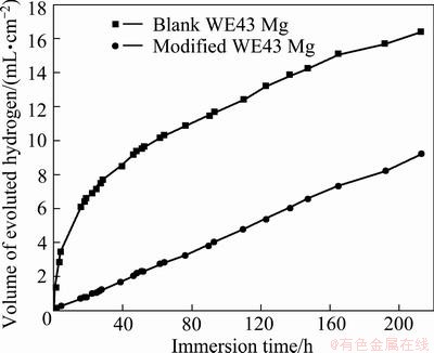
Fig. 3 Hydrogen evolution volume of phosphating coated WE43 alloy
3.3 Electrochemical test
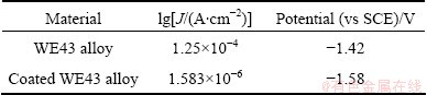
Figure 4 presents the electrochemical polarization curves of different samples immersed in SBF solutions at 37 ��C. The average electrochemical parameters are also shown in Table 1. The results show that the corrosion potential changes from -1.42 V for the untreated sample to -1.58 V for the coated sample, while the current density changes from 1.583��10-6 to 1.125��10-4 A/cm2, respectively, which suggests that the phosphating coated WE43 magnesium alloy has a better corrosion resistance compared with the uncoated alloy.
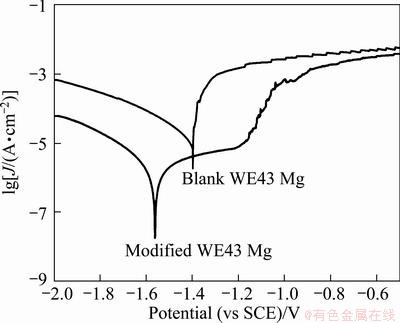
Fig. 4 Potentiodynamic polarization curves of WE43 alloy with and without phosphating in SBF solution at 37 ��C
Table 1 Average electrochemical parameters of potentio- dynamic polarization of WE43 alloy with and without phosphating in SBF solution at 37 ��C
3.4 Biocompatibility evaluation
Figure 5 illustrates the viabilities of L-929 cells expressed as a percentage of the viability of cells cultured in the negative control and the phosphating coated WE43 alloy extraction medium solutions for different periods (1, 2 and 4 d). It can be seen that the coated group has a better cell viability than the control. At the three time points, the viabilities of the three coated samples are all above 80% while the uncoated WE43 alloy is about 30%, as shown in Table 2. It may be because of the good biocompatibility of phytic acid and fluorine as fluorine is a normal element in the body. KIM et al [23] and HAUGEN et al [24] proved that the cell viability can be promoted by fluorine addition. In addition, phytic acid is widely present in nature (plants, animals and soils), mainly as the calcium, magnesium and potassium mixed salts (also called phytines), and it has important biological activities and wide applications. Since the last century, it has been the subject of investigation for many scientists in different fields, such as enhanced immunity [25] and anticancer [26,27]. Therefore, the present results indicate that this surface modification method can promote the cell viability of magnesium alloys.
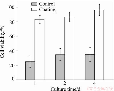
Fig. 5 L-929 cell viability in control and phosphating coated WE43 alloy extraction solutions after 1, 2 and 4 d of culture
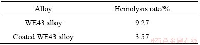
For the hemolysis test, compared with the control group, the hemolysis rate of the coated WE43 alloy changed from 9.27% to approximate 3.57%, as shown in Table 3, which shows that after the phosphating coating, the blood compatibility of the WE43 magnesium alloy can be improved significantly.
Table 2 L-929 cell viability in uncoated and coated WE43 alloy extraction solutions after 1, 2 and 4 d of culture
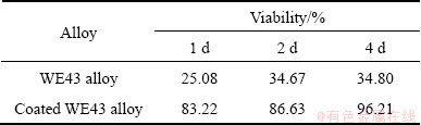
Table 3 Hemolysis rate of WE43 alloy with and without phosphating coating
4 Conclusions
1) The results show that the phosphating solution without any poisonous composition to the body can improve the corrosion resistance of WE43 alloy in the SBF, compared with the untreated alloy.
2) For cell viability study, the viability of the surface modified alloy is above 80% while that of the control groups is about 30%, which illustrates that the phosphating coated WE43 alloy can meet the basic requirement for biomaterials, meanwhile, the blood compatibility results also show that the hemolysis rate of the phosphating coated alloy is decreased down to 3.57%, which also means that the coated alloy can meet the requirement for biomaterials.
3) Phosphating treatment should be a promising technique to improve both corrosion resistance and biocompatibility of the biodegradable WE43 magnesium alloy.
References
[1] AI H J, HONG Y S, YANG K, ZHANG G D, HUANG J J, HAO Y Q. The role of bone induction of a biodegradable magnesium alloy [J]. Acta Metall Sin, 2008, 44: 1035-1041.
[2] DUYGULU O, KAYA R A, OKTAY G, KAYA A A. Investigation on the potential of magnesium alloy AZ31 as a bone implant [J]. Mater Sci Forum, 2007, 546-549: 421-424.
[3] YANG J X, CUI F Z, YIN Q S, ZHANG Y, ZHANG T, WANG X M. Characterization and degradation study of calcium phosphate coating on magnesium alloy bone implant in vitro [J]. Ieee T Plasma Sci, 2009, 37: 1161-1168.
[4] ZHANG E L, XU L P, YU G N, PAN F, YANG K. In vivo evaluation of biodegradable magnesium alloy bone implant in the first 6 months implantation [J]. J Biomed Mater Res A, 2009, 90: 882-893.
[5] EGGEBRECHT H, RODERMANN J, HUNOLD P, SCHMERMUND A, BOSE D, HAUDE M, ERBEL R. Novel magnetic resonance-compatible coronary stent��The absorbable magnesium-alloy stent [J].Circulation, 2005, 112: E303-E304.
[6] GHIMIRE G, SPIRO J R, KHARBANDA R K, ROUGHTON M, BARLIS P, MASON M, ILSLEY C, DI MARIO C, ERBEL R, WAKSMAN R, DALBY M C D. Evidence for the return of coronary vasoreactivity following absorption of a bioabsorbable magnesium alloy coronary stent [J]. Euro Intervention, 2009, 4(4): 481-484.
[7] MAENG M, JENSEN L O, THUESEN L. Constrictive vascular remodeling after implantation of a bioabsorbable magnesium alloy stent in porcine coronary arteries [J]. J Am Coll Cardiol, 2008, 51: B91.
[8] WAKSMAN R, PAKALA R, HELLINGA D, BAFFOUR R, KUCHULAKANTI P, SEABRON R, TIO F. Effect of bioabsorbable magnesium alloy stent on neointimal formation in a porcine coronary model [J]. Catheterization and Cardiovascular Interventions, 2006, 68(4): 607-617.
[9] ZHENG Y F, GU X N, ZHENG W, CHENG Y. A study on alkaline heat treated Mg-Ca alloy for the control of the biocorrosion rate [J]. Acta Biomater, 2009, 5: 2790-2799.
[10] CHU P K, LIU C L, XIN Y C, TANG G Y. Influence of heat treatment on degradation behavior of bio-degradable die-cast AZ63 magnesium alloy in simulated body fluid [J]. Mat Sci Eng A, 2007, 456: 350-357.
[11] ZHENG Y F, GU X N, LI N, ZHOU W R, ZHAO X, CAI Q Z, RUAN L Q. Corrosion resistance and surface biocompatibility of a microarc oxidation coating on a Mg-Ca alloy [J]. Acta Biomater, 2011, 7: 1880-1889.
[12] SONG Y W, SHAN D Y, HAN E H. Electrodeposition of hydroxyapatite coating on AZ91D magnesium alloy for biomaterial application [J]. Mater Lett, 2008, 62: 3276-3279.
[13] LIAN J S, LI G Y, NIU L Y, JIANG Z H, JIANG Q. Growth of zinc phosphate coatings on AZ91D magnesium alloy [J]. Surf Coat Tech, 2006, 201: 1814-1820.
[14] ZHANG E L, XU L P, PAN F, YU G N, YANG L, YANG K. In vitro and in vivo evaluation of the surface bioactivity of a calcium phosphate coated magnesium alloy [J]. Biomaterials, 2009, 30: 1512-1523.
[15] JIN G, CUI X F, LI Q F, LI Y, WANG F H, DING M H. Microstructure and corrosion resistance of phytic acid conversion coatings for magnesium alloy [J]. Appl Surf Sci, 2008, 255: 2098-2103.
[16] LIU J R, GUO Y N, HUANG W D. Study on the corrosion resistance of phytic acid conversion coating for magnesium alloys [J]. Surf Coat Tech, 2006, 201: 1536-1541.
[17] CUI X F, LI Y, LI Q F, JIN G, DING M H, WANG F H. Influence of phytic acid concentration on performance of phytic acid conversion coatings on the AZ91D magnesium alloy [J]. Mater Chem Phys, 2008, 111: 503-507.
[18] GAO H F, ZHANG S T, LIU C L, XU Q, LI J, LI Y. Effect of pH on phytic acid conversion coating on AZ31B magnesium alloy [C]//Proceedings of the 7th National Conference on Chinese Functional Materials and Applications. Changsha: Instrument Materials Socienty of China Instrument and Control Society, 2010: 414-1419.
[19] LIANG C H, ZHENG R F, HUANG N B, XU L S. Conversion coating treatment for AZ31 magnesium alloys by a phytic acid bath [J]. J Appl Electrochem, 2009, 39: 1857-1862.
[20] XU L, ZHANG E, YANG K. Phosphating treatment and corrosion properties of Mg-Mn-Zn alloy for biomedical application [J]. J Mater Sci Mater Med, 2009, 20(4): 859-867.
[21] KOUISNI L, AZZI M, ZERTOUBI M, DALARD F, MAXIMOVITCH S. Phosphate coatings on magnesium alloy AM60 Part 1: Study of the formation and the growth of zinc phosphate films [J]. Surf Coat Tech, 2004, 185(1): 58-67.
[22] SAMMARTANO S, CREA P, de ROBERTIS A, de STEFANO C. Speciation of phytate ion in aqueous solution. Sequestration of magnesium and calcium by phytate at different temperatures and ionic strengths, in NaCl(aq) [J]. Biophys Chem, 2006, 124: 18-26.
[23] KIM H E, LEE S H, KIM H W, KONG Y M, LEE S H, CHANG Y I. Fluoride coatings on orthodontic wire for controlled release of fluorine ion [J]. J Biomed Mater Res B, 2005, 75: 200-204.
[24] HAUGEN H J, TIAINEN H, MONJO M, KNYCHALA J, NILSEN O, LYNGSTADAAS S P, ELLINGSEN J E. The effect of fluoride surface modification of ceramic TiO2 on the surface properties and biological response of osteoblastic cells in vitro [J]. Biomed Mater, 2011(6): 1-12.
[25] SHAMSUDDIN A M, VUCENIK I. Mammary tumor inhibition by IP6: A review [J]. Anticancer Res, 1999, 19(5A): 3671-3674.
[26] BATEN A, ULLAH A, TOMAZIC J V, SHAMSUDDIN A M. Inositol-phosphate-induced enhancement of natural killer cell activity correlates with tumor suppression [J]. Carcinogenesis, 1989, 10(9): 1595-1598.
[27] GRAF E, EATON J W. Antioxidant functions of phytic acid [J]. Free Radical Bio Med, 1990, 8(1): 61-69.
������WE43þ�Ͻ���������������Լ���ʴ����
Ҷ�ɺ�1����͢�1,2��֣���1,3��������2�������4
1. ������ѧ ǰ�ؽ���ѧ���о�Ժ ����ҽ�ò�������֯�����о����ģ����� 100871��
2. ����ҽѧԺ ��Ϣ�빤��ѧԺ������ 325035��
3. ������ѧ ��ѧԺ �Ƚ�������������ϵ������ 100871��
4. ��ݸ�˰��Ƽ��ɷ�����˾����ݸ 523000
ժ Ҫ��ͨ�����ݷ�ʽ�Ʊ�������WE43þ�Ͻ�����ɨ��羵(SEM)��X��������(XRD)�ȷ����������κ�þ�Ͻ�ı����۽ṹ��ͨ���绯ѧ�������Բ��ϵĿ���ʴ���ܣ�ͬʱ������MTT������Ѫ�����鷨�Ƚ������κ�δ����þ�Ͻ���ϵ����������ԡ�����������������ܴ���ȵ����WE43þ�Ͻ����ʴ�Լ����������ԡ�
�ؼ��ʣ�þ�Ͻ������Σ���ʴ���ܣ�����������
(Edited by Xiang-qun LI)
Foundation item: Project (2011AA030103) supported by the National High-tech Research Program of China; Project (201001C0104669453) supported by the Guangdong Innovation R&D Team Project, China
Corresponding author: Ting-fei XI; Tel/Fax: +86-10-62753404; E-mail: xitingfei@tom.com
DOI: 10.1016/S1003-6326(13)62558-3

