
��TGF-��1������Ϳ�����Ѷ�MG63ϸ�����ܵ�Ӱ��
������1, 2������1����Сƽ1��������2���κ�2����ͦ2
(1. ���ϴ�ѧ ������ҽԺ������ ��ɳ��410013��
2. ���ϴ�ѧ ��ĩұ������ص�ʵ���ң����� ��ɳ��410083)
ժҪ���ø����黯�����ۺϽ������Ʊ���������������������TGF-��1�������MG63ϸ����ֳ�ͷֻ���Ӱ�죬���Ż�TGF-��1����Ũ�ȣ��÷�ĩע����μ����Ʊ�60%��϶�Ⱦ�����ͨ�ṹ����ѣ���Ϳ�����ڶ���ѱ����϶���Ʊ���TGF-��1������Ϳ�㣬MTT������Ϳ���L929ϸ�����ԣ�ELISA������Ϳ���ҩ�ﶯ�����ԣ��������۸�Ϳ���MG63ϸ����𤸽����ֳ�ͷֻ���Ӱ�졣�о����������TGF-��1Ũ��Ӱ��MG63ϸ�����ܣ���0.025~2.500 mg/g��Χ�ڣ�TGF-��1��MG63ϸ������ֳ�ͷֻ����óʼ�����ЧӦ��ϵ, Ũ��Ϊ2.5 mg/gʱ����MG63ϸ���ֻ��������ţ���TGF-��1Ũ��Ϊ25 mg/gʱ����MG63ϸ����ֳ���������ã���TGF-��1������Ϳ��������ϸ�����ԣ�Ϳ����л����ܣ���ά��12d����Ϳ��������MG63ϸ����𤸽����ֳ�� �ֻ���
�ؼ��ʣ�
���������TGF-��1��Ϳ�����ɹ�ϸ������ֳ���ֻ���
��ͼ����ţ�R783.1 ���ױ�־�룺A ���±�ţ�1672-7207(2011)08-2266-08
Effects of TGF-��1 gelatin microspheres coated on
porous titanium on function of MG63 cells
CHEN Liang-jian1, 2, WANG Rui-fang1, GUO Xiao-ping1, LI Yi-min2, HE Hao2, LI Ting2
(1. Third Xiangya Hospital, Central South University, Changsha 410013, China;
2. State Key Laboratory of Powder Metallurgy, Central South University, Changsha 410083, China)
Abstract: Gelatin microspheres were made by improved emulsified cold condensation method. Effects of TGF-��1 gelatin microspheres on the proliferation and differentiation of MG63 cells were investigated in vitro, as well as the optimal TGF-��1 concentration. Porous titanium with a porosity of 60% and an inter-connected structure was prepared using a powder injection molding method. A coating of TGF-��1 loaded gelatin microspheres was applied in the pores of the porous titanium surface. MTT assay method was used to evaluate the cytotoxicity of the coating to L929 cells. The pharmacokinetics of the coating was tested with TGF-��1 ELISA kit. In vitro, the effect of the coating on adhesion, proliferation and differentiation of MG63 cells was investigated. The results show that the TGF-��1 concentration affects the function of MG63 cells. In vitro, the proliferation and differentiation of MG63 cells show a positive dose-effect relationship at TGF-��1 concentrations of 0.025-2.500 mg/g. TGF-��1 concentration of 2.500 mg/g is in favor of the differentiation, while the concentration of 25 mg/g inhibites cells�� proliferation. Porous titanium samples coated with TGF-��1 gelatin microspheres are nontoxic to L929 cells and are able to maintain a slow-release state during 12 d. The porous titanium implants coated with TGF-��1 loaded gelatin microspheres benefits MG63 cells adhesion, proliferation and differentiation on the 3th, 7th, 14th slot.
Key words: porous titanium; TGF-��1 loaded gelatin microspheres coating; MG63 cell; proliferation; differentiation
��ֲ�������Ϊ����ȱʧ���������������е�����ֲ���Ϊȫ�����ͣ��д����²��㣺δ�ṩ����֯����Ľṹ�������������γ�ʱ�䳤(��3~6��)�������ȶ��Եͣ���ֲ�����Ϳ������ֲ��������ƻ�ʧЧ�ȡ������ֲ�����ṩ���ʳ�����ֲ��Ľṹ��ʵ������̶�������ѧ�����������ƥ�䣬��������ֲ����о��ȵ㡣��ǰ����ֲ������о��������ڸ�����ֲ����ϸ�����������ٽ����׳������¹����ɣ�ȱ����ֲ������Χϸ������֯����㻥��������������ֲ����������Ա��Χϸ����������ǿ���ɹ��źţ��ӿ���ֲ����Χ����֯�ĸĽ������ϣ��������������ֲ����ڵ��ȶ��Ժͳɹ��ʡ������о�֤ʵ�ֲ�Ӧ���������ӿ��Դٽ��������ϸ����ɹ�ϸ���ֻ������������ø���Ժ����ӹ�����mRNA�ı���[1-2]������������ֱ�Ӿֲ�Ӧ�ô�����ɢ�Ͽ졢�ױ�����ø�ֽ⡢��˥�ڶ̵�ȱ��[3]������������ЧŨ�Ⱥ�����ʱ���ά���Ǿֲ�Ӧ�õ�ǰ�ᡣ������������������Ϊ�������ӵĻ���ϵͳ��ͨ������ϸ��ʵ���о���TGF-��1����������Ϳ��Ķ���������Գɹ�ϸ������Ӱ�죬���Ż���������TGF-��1����Ũ�ȡ�
1 ���Ϻͷ���
1.1 ��TGF-��1������Ϳ�����ѵ��Ʊ�
1.1.1 ����ѵ��Ʊ�
�÷�ĩע����μ����Ʊ���϶��Ϊ60%����Ϊ50~300 ��m������ͨ�ṹ�����[4]���ñ�ͪ����ˮ�Ҵ���ȥ����ˮ��������ϴ��10 min�����¶�Ϊ60 ��ĺ�����и�����¸�ѹ�������á�
1.1.2 ��TGF-��1��������Ʊ�
�ø����黯�����ۺϽ������Ʊ�����Ϊ10~40 ��m������������[1]��ÿ1 mg�������зֱ����0.5��5��50��500 mg/L��TGF-��1��Һ��5 ��L����4 �棬pH=7.4�����£�����24 h��������ͺ�����(1 200~ 1 500 r/min)15 min, ˫��ˮϴ��2�Σ����ɣ���-60���������档
1.1.3 ��TGF-��1������Ϳ�����ѵ��Ʊ�
����������Ϊ5%��������Һ���ݶ���ѣ���ѹ�����´���10 min��50 �����6 h������ˮ�Ҵ�������������Ϊ20 mg/mL TGF-��1��������ҺͿ�����ѱ��棬����������Ϊ2.5%�����ȩ��Һ��������30 min������ˮ�Ҵ���ϴ3�Σ�ÿ��10 min���䶳�����-60���������档
1.2 ϸ������ʵ��(MTT��)
��������0.2 g/mL�ı��������ں�10%СţѪ���RPMI 1640�������У���ϸ���������н���72 h ���ռ�����Һ��4 �汣�档��L929ϸ����Һ��2.5 ��104��/mL������96��ϸ��������(200 ��L/��)������24 h��������ȡŨ�ȷֱ�Ϊ100%��50%��10%(�����������ͬ)�Ľ���Һ����ԭ����Һ��ÿ����6��ƽ�пס����Զ�����Ϊ��10%СţѪ�������RPMI1640�����������Զ��������10 ��LŨ��Ϊ8 mol/L�ı�����Һ����������3 d������������(MTT)��Һ����������4 h�������壬�����������������ø���������ڲ���490 nm�²�������ȡ�
1.3 ������ҩʵ��
���ѡ����TGF-��1Ũ�ȷֱ�Ϊ0.025��0.25��2.5��25 mg/g������Ϳ������������3����������24��ϸ�������壬����2 mL��1640�����������������24 h��ȡ����Һ��4 �汣�棬��Ϊ�ͷ�24 h�Ĵ������Һ����ͬ��������ȡ2��4��6��8��10��12 d�Ľ���Һ��4 �汣�档��TGF-��1 ELISA�Լ��еIJ���˵������ø����������450 nm�����²ⶨ����Һ����ȣ���TGF-��1�����߶��գ������ʱ�������TGF-��1Ũ�ȣ�����TGF-��1�ͷ����ߡ�
1.4 �Ż���������TGF-��1Ũ��
���ز�ͬŨ��TGF-��1��������MG63ϸ������������ͨ�������TGF-��1�������MG63ϸ������ֳ�ͷֻ�Ӱ�죬�Ż���Ũ�ȡ�
1.4.1 ϸ����ֳ���
��0.25%��ø����MG63ϸ��, �ú�12%̥ţѪ���1640����������ϸ��Ũ��Ϊ5��104��/mL��ϸ����Һ��������24�װ��ڣ�����24 h��ϸ�����ڣ������Ƶ���TGF-��1Ũ�ȷֱ�Ϊ0.025��0.250��2.500��25.000 mg/g������2 mg�������鲻���������ӵ���2 mg����3�����ף��ֱ�����3��7��14 d����SunBioTMAm-Blueϸ����ֳ����Լ���Լ�˵����������ڲ���490 nm�·ֹ���ø���Dzⶨ����������Ȳ���¼�����ձ����ߣ�����ϸ����ֳ����
1.4.2 ϸ���ֻ����
ȡ�������Ĺ��е�3��7��14 �������Һ, ����������ø(ALP)�ⶨ�Լ���˵����(�Ͼ��������﹤�̹�������һ����)���в�������ø�������Dzⶨ��������Ȳ���¼�����ձ����ߣ�����ALP�Ļ��ԡ�
1.5 ��TGF-��1������Ϳ��Ķ����������MG63ϸ����Ӱ��
1.5.1 ϸ����ֳ������
����3��ʵ�飬ʵ����Ϊ��2.5 mg/g TGF-��1������Ϳ�����ѣ�������Ϊֱ����100 ��L 10 mg/L TGF-��1��Һ��������ѱ��棬δ������Ϊ����TGF-��1�������������3�����ף���0.25%��ø����MG63ϸ������10%СţѪ���1640������������Ũ��Ϊ4.0��104��/mL����Һ��ȡ2 mLϸ����Һ�����ڷ���3��������24�����������������ֱ��ڵ�3��7��14�죬��SunBioTMAm-Blueϸ����ֳ����Լ���Լ�˵����������ڲ���490 nm�·ֹ���ø���Dzⶨ��������Ȳ���¼�����ձ����ߣ�����ϸ����ֳ����
1.5.2 𤸽��ò���
������MG63ϸ�������������ֱ��ڵ�7�͵�14����ȥ����Һ���ټ���4 ��Ԥ���2.5%���ȩ�̶�����4 �汣�档�þƾ��ݶ���ˮ���ٽ�������ɨ��羵���MG63ϸ���ڶ���ѱ����𤸽��ò��
1.5.3 ϸ���ֻ�����
��������ø(ALP)��������MG63ϸ����������3��7��14 d���ռ���������Һ����ALP�ⶨ�Լ���˵����(�Ͼ��������﹤�̹�������һ����)���в�������ø�������Dzⶨ��������Ȳ���¼�����ձ����ߣ�����ALP�Ļ��ԡ�
�Ǹ���(BGP)��������MG63ϸ����������3��7��14 d���ռ���������Һ������[125I]�Ǹ��ط������߷���ҩ��(�����ն�ΰҵ����Ƽ�����˾)˵������в������÷����Լ���Dzⶨ�������Լ���ֵ����¼�����ձ����ߣ�����BGP���ԡ�
1.6 ͳ�Ʒ���
ѡ��SpSS13.0ͳ����������������ƽ��ֵ(![]() )������(s)��ʾ(
)������(s)��ʾ(![]() ��s)�����õ����ط��������P��0.05ʱ�������������ԡ�
��s)�����õ����ط��������P��0.05ʱ�������������ԡ�
2 ���
2.1 ��TGF-��1������Ϳ�����ѱ�����̬
��TGF-��1��������Ϳ������SEMͼ��ͼ1��2��ʾ���ɼ�����TGF-��1�����������Բ����, �����Ͼ��ȣ�����⻬��δ�������ơ�����ѵı���Ϳ�϶���ϳ�����������Ϳ�㡣
2.2 ϸ����������
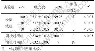
ʵ��ϸ��ΪL929 ϸ����3��ʵ����ֱ�ΪŨ��100%��50%��10%�Ľ���Һ�����Զ���������Զ����顣��������3 d ����MTT ���������ϸ�����ԣ���490 nm ø��������Dzⶨ����ȣ�������ȼ��������ֳ��(R)��R=De/Dn��100%��ʽ�У�DeΪ�����������ȣ�DnΪ���Զ����������ȡ���R����ϸ�����Էּ�(CTS)������1���ɱ�1 ������3 ��Ũ�ȵĽ���Һ��R ֵ�����Զ�����Ƚϲ����������(p��0.05)��CTS�ּ���Ϊ0~������������Ϳ������������ϸ�����ԣ����ϡ�ҽ����е����ѧ���۹��ұ�GB/T 16886.1��2001���жԲ���ϸ�����Ե�Ҫ��
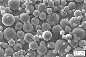
ͼ1 ��TGF-��1������SEMͼ
Fig.1 SEM image of TGF-��1 loaded gelatin microspheres
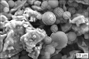
ͼ2 ��TGF-��1������Ϳ������SEMͼ
Fig.2 SEM image of porous titanium coated with TGF-��1 loaded gelatin microspheres
��1 ������Ϳ�����Ѷ�L929ϸ�����Խ��
Table 1 Results of cytotoxicity tests of porous titanium coated with TGF-��1 loaded gelatin microspheres with L929 cells
2.3 �����ͷ�ʵ��
�������ӵ���ɢ�ٶȺ�������ϵĽ����ٶȣ��������ڼ�����������ȫ����Լ��2�£���ҺpH=7.35~7.45��������������������ò���Ѫ���ϸ������ҺΪ�ͷŽ��ʣ���TGF-��1 ELISA�Լ��еIJ���˵������ͬTGF-��1Ũ�ȵ�����������Ϳ�����������ͷ�������ͼ3��ʾ����ͼ3�ɿ����������ͷ��ٶȽϿ죬24 h�ڴﵽ20%~30%�����ͷ�������TGF-��1��Ũ����أ���3��ʱ�ۼ��ͷ�����40%����ʱ�����ӣ��ͷ��ٶȼ�������8��ʱ��4��Ũ�������ͷ����ʽӽ�һ�£���12��ʱ�ۼ��ͷ���Ϊ93%��
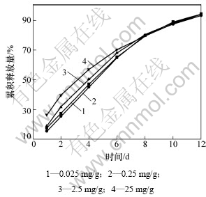
ͼ3 ��ͬTGF-��1����������������Ϳ�����������ͷ�����
Fig.3 Release profile of porous titanium coated with different concentration TGF-��1 loaded gelatin microspheres
2.4 ��TGF-��1����������Ũ�ȵ��Ż�
2.4.1 TGF-��1Ũ�ȶ�ϸ����ֳӰ��
MG63ϸ������TGF-��1����������3��7��14 d��SunBioTMAm-Blueϸ����ֳ����Լ���Լ�˵���������MG63ϸ���ڲ�ͬŨ��TGF-��1������3��7��14 dʱ��ֳ�ıȽ���ͼ4��ʾ����ͼ4�ɿ�����ͬһʱ��㲻ͬŨ��TGF-��1�������MG63ϸ������ֳӰ�첻ͬ��TGF-��1Ũ����0.025~0.25 mg/g��Χ�ڣ�TGF-��1��Ũ����MG63ϸ����ֳ�ʼ�����ЧӦ��ϵ����TGF-��1Ũ��Ϊ25 mg/gʱ��TGF-��1��MG63ϸ����ֳ���������á�
2.4.2 TGF-��1Ũ�ȶ�ϸ���ֻ�Ӱ��
MG63ϸ������TGF-��1����������3��7��14 d��ALP�Լ��в�����MG63ϸ���ڲ�ͬŨ��TGF-��1������3��7��14 dʱALP�Ļ�����ͼ5��ʾ����ͼ5�ɿ�������0.025~2.5 mg/g��Χ�ڣ�TGF-��1Ũ�ȶ�MG63ϸ���ֻ��ʼ�����ЧӦ��ϵ����TGF-��1Ũ��Ϊ2.5 mg/gʱ���������MG63ϸ���ֻ��ٽ�������������4�顣
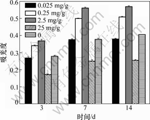
ͼ4 MG63ϸ���ڲ�ͬ����TGF-��1������3��7��14 dʱ��ֳ�ıȽ�
Fig.4 Comparison of cell proliferation of MG63 and different concentration of TGF-��1 after cultured 4, 7 and 14 d
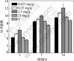
ͼ5 MG63ϸ���ڲ�ͬŨ��TGF-��1������3��7��14 dʱALP�Ļ���
Fig.5 ALP activity of MG63 cells and different concentration of TGF-��1 after cultured 3, 7 and 14 d
2.5 ��TGF-��1������Ϳ��Գɹ�ϸ�����ܵ�Ӱ��
2.5.1 ϸ����ֳ
3��������MG63ϸ����������3��7��14 d����SunBioTMAm-Blue�Լ��в�����3������������MG63ϸ����������3��7��14 d��ϸ����ֳ�ıȽ���ͼ6��ʾ���ɼ���������3��7 dʱ��ʵ����ɹ�ϸ��������࣬��������Σ�δ���������٣���14��ʱ��ʵ�����������2�飬����������δ������ϸ����ֳ��������������ԡ�
2.5.2 𤸽��ò
3��������MG63ϸ����������7��14 d��ɨ��羵���MG63ϸ����������𤸽ϸ����������ò��3��������MG63ϸ����������7��14 d �ı�����ò��ͼ7��ʾ���ɼ���7 dʱ��ʵ������������ϸ������Խ϶࣬ϸ����̬������α��࣬ϸ�������ڿ�϶��Ե�������϶��Ǩ������(ͼ7(a))����������������𤸽��MG63ϸ��������ʵ�����٣�ϸ����չ���ã�ϸ�����С��϶�γ�ϸ����(ͼ7(c))��δ��������������𤸽ϸ����ʵ����Ͷ�������٣�ϸ�������Σ�α����(ͼ7(e))��14 dʱ��3�����������ϸ�����������ã�ϸ����ʲ��������Σ�ʵ����ϸ��α����������棬��չ���ã�ϸ��������϶��ں͵ױ�������ϸ����Խ��϶���ӳ�Ƭ���ʵ���״�ṹ(ͼ7(b)����������������𤸽ϸ������ò��ʵ�������ƣ�ϸ����Խ��϶���ӳ�Ƭ����(ͼ7(d))��δ������ϸ�������ױںͿױ���������δ�γɵ���״�ṹ(ͼ7(f))������������ɿ�����������MG63ϸ����������7 dʱ��ʵ������������𤸽ϸ���������Զ��ڶ������δ�����飬ϸ����̬������α��ࣻ14 dʱ��ʵ����Ͷ�������������𤸽ϸ�����������Զ���δ�����飬ϸ����Խ��϶�������ӳ�Ƭ���ʵ���״�ṹ𤸽�ڿ�϶��ں͵ױڣ�δ��������������𤸽��ϸ�����ص㡣
ͼ6 3������������MG63ϸ����������3��7��14 d��ϸ����ֳ�ıȽ�
Fig.6 Comparison of cell proliferation of MG63 and three groups porous titanium samples after cultured 3, 7 and 14 d
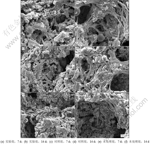
ͼ7 3��������MG63ϸ����������7��14 d �ı�����ò
Fig.7 Surface morphologies of MG63 cells cultured for 7 d and 14 d on three groups porous titanium samples
2.5.3 ϸ���ֻ�
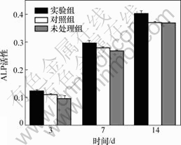
(1) ALP�ļ�⡣3��������MG63ϸ����������3��7��14 d����ALP�Լ��м������Һ��ALP���ԡ�3��������MG63ϸ����������3��7��14 d��ALP�Ļ�����ͼ8��ʾ����ͼ8�ɼ�������3��7 dʱ��ʵ�������������ALP���Ծ�����δ�������ALP���ԣ���δ������Ƚϲ�����������(P��0.05)����ʵ������������Ƚϲ�����������(P��0.05)��14 dʱ��ʵ����ALP���Ը��ڶ�����ALP���ԣ���������ALP���Ը���δ������ALP���ԣ����Ƚϲ�����������(P��0.05)��
(2) �Ǹ��ؼ�⡣3��������MG63ϸ����������3��7��14 d���ùǸ����Լ��м������Һ�йǸ���(BGP) ���ԣ������ͼ9��ʾ����ͼ9�ɼ�������3��7 dʱ��ʵ����Ͷ������BGP���Ծ�����δ�������BGP���ԣ���δ�������BGP���ԱȽϲ�����������(P��0.05)����ʵ�����������������������(P��0.05)��14 dʱ��ʵ�����BGP���Ը��ڶ�����BGP���ԣ�������BGP���Ը���δ������BGP���ԣ����Ƚϲ�����������(P��0.05)��������ALP��BGP�ļ������֪�������������TGF-��1������������MG63ϸ���ķֻ�����TGF-��1������Ϳ���MG63ϸ������������ֱ����TGF-��1��Һ�顣
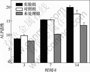
ͼ8 3��������MG63ϸ����������3��7��14 d��ALP�Ļ���
Fig.8 ALP activity of MG63 cells and three groups porous titanium samples after cultured 3, 7 and 14 d
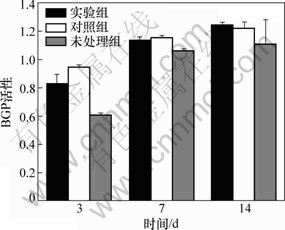
ͼ9 3��������MG63ϸ����������3��7��14 d��BGP����
Fig.9 BGP activity of MG63 cells and three groups porous titanium samples after cultured 3, 7 and 14 d
3 ����
�Ѻ��ѺϽ������������ѧ���ܺ����������ԣ��ѱ��㷺Ӧ���ڹǿƵ��˹��ؽڼ��弰����ֲ�塣��������Բ��ֲ����ֲ����γɹ�����ʱ��һ����3~6 �¡���������ֲ������������ԣ��ӿ�������γ���������ֲ����о��ȵ㡣�ֲ�Ӧ�ôٹ���֯�γɵ�������������ֲ�������ԵIJ���֮һ�������о�֤ʵ�ڹ�������ϱ������������������ӿ��Դٽ��ɹ�ϸ���ķֻ������ʷ��ڼ��ƻ��������ٽ���˿��������(��IGF-1)����ǿ��ϸ�����Ե�����(��TGF-��1)���յ��ɹǵ�����(��BMPS ��)[1]��TGF-��1�дٳɹǺͳ��������ã��ı��Ĥ��ϸ�����ɣ��յ��Ĺ��γ�[5-6]����Դ���������Ӵ����ȶ��Ե͡�����Ĥ���Բ��˥�ڶ̡��ֲ�ֱ��Ӧ�������ںܿ챻ϡ�ͺͷֽ�Ȳ��㡣ҩ�ﻺ��ϵͳ���л���ҩ��������͡��ڱ�֤ҩ���������õ�ǰ���¼��ٸ�ҩ����������ҩ�ﶾ�Ե��ŵ�[7]���ֲ����û���ϵͳ�������������ܽ���������㣬��ѡ�õ�������Ϻͺ���������������Ũ��Ŀǰ�д����顣�������Ʊ�����ϵͳ�IJ����о����ᡢ�������Ǿ��ǵȣ������������ڽ������Ϊ���ᣬ���¾ֲ�������֢��Ӧ[8-9]�����о�ѡ������Ϊ���Ͳ��ϣ���������֯�����Ժã�����Ȼ���⣬������ﲻ������֯��֢��Ӧ[10]�����ɰ����������ཻ���γɵ�ֱ���ۺ���ܽ��ж��ֱ������μ���Ӧ���Һ��о�����-�ʰ���-�춬����(RGD)������Զ��ģ�����ϸ�����������ϣ�������ϸ������𤸽����ʶ ��[11]����������Ϊ���Ľṹ�����������������У�������������γ���������ȶ����ĵ���Ȼ���Ӷ������������ӻ��ԣ���������ҩ���ͷ��ٶ���Ի������ܴﵽ���͵�Ҫ��[12-13]�����������Ѳ��ø����黯�����ۺϽ������Ż����������Ʊ��� ��[14]�����о��ڴ˻������Ż�������TGF-��1Ũ�ȣ��о���TGF-��1������Ϳ�����������Գɹ�ϸ����Ӱ�졣
��ֲ��ֲ�����10~12 d�ɹ�ϸ������ֲ��ǽ��濪ʼ�ֻ��ɹǣ�12~18 d�ɹ�ϸ��������ǿ���ǽ��濪ʼ�ǸĽ�[15]������ֲ�����������ﻯ���ԣ��ٽ��������ڳɹ�ϸ��ļ���ͷֻ��������ڹ������γɡ�TGF-��1�ٽ��ѱ�ļ�����ijɹ�ϸ���ϳɢ��ͽ�ԭ����ά��ϵ��ף����������ƹ�ϸ���Ļ���̼��ɹ�ĸϸ������ֳ�ͻ������[10, 16]���ʼ��������Դٽ���Դ�Լ����ϸ�������Ƿֻ���TGF-��1Ũ�ȶ�����ϸ���дٽ��ͷֻ���˫�ص�������[17]������ֲ�������TGF-��1���б�����ԣ��������ڴٽ��������������ϳ��������ڹ������γɣ���Ŀǰ�ֲ�Ӧ���������ӵ���ѱ�¶ʱ������Ũ�������ۡ�һ����Ϊ��Ũ��TGF-��1�ٽ�ϸ����ֳ������Ũ������ϸ����ֳ[18]�����о�����TGF-��1Ũ����0.01~1.00 mg/L��Χ�ڣ���������ӶԳɹ�ϸ������ֳ����Ҳ��ǿ������Ũ��Ϊ1~10 mg/Lʱ����ϸ����ֳ���ò����ԣ�����Ũ�ȴ���10 mg/Lʱ����Ϊ��ϸ����ֳ����������[19]����Ҳ���о���ΪTGF-��1�ڵ�Ũ��(С��0.01 mg/L)ʱ�ɴٽ��ɹ�ϸ������ֳ������0.01 mg/Lʱ����Ũ�ȵ����ӣ�����ֳ�������Լ���[4]�����о�����ʵ�鷢�֣���TGF-��1��������MG63ϸ������������TGF-��1Ũ��Ϊ0.025 mg/g���������MG63ϸ����ֳ���������ã�25 mg/gʱ��MG63ϸ����ֳ���������ã�Ũ����0.25~2.5 mg/g��Χʱ����MG63ϸ����ֳ�ͷֻ����дٽ����ã���ϸ����ֳ�ʼ�����ЧӦ��ϵ��TGF-��1Ũ��Ϊ2.5 mg/g���������ϸ����ֳ�������š�
���ϱ�����ò�ʹֲڶȡ����滯ѧ��ɡ���ˮ�ԡ������ܺͱ�����Ӱ��ϸ��𤸽���ֲڵı���ṹ�����õı��滯ѧ�������ˮ�ԡ��߱����ܺ��ʺϵı����������ڳɹ�ϸ����𤸽[20-21]�����о�������TGF-��1������Ϳ���ֱ��Ϳ��TGF-��1�Ķ����������MG63ϸ����������7��14 d����������𤸽��������ϸ����̬����չ״��������δ�����Ķ��������������7 dʱ��ʵ������������𤸽��ϸ���������Զ��ڶ����顣ʵ�����MG63ϸ��𤸽����ֳ��������B��δ�����飬���������������йأ��������ɰ����������ཻ���γɵ�ֱ���ۺ����������ϸ�����������������ϵ�������Զ���(RGD)��������ϸ������𤸽����ʶ��[11]����ϸ��𤸽�����������϶�ڳ�����TGF-��1�����ɳ����ͷ�TGF-��1����ά��TGF-��1Ũ������ȶ���������ϸ������������ļ�����ٽ�ϸ���ϳɷ���ϸ������ʣ�Ϊϸ��𤸽�ṩ��������3��7 dʱ��������������MG63ϸ������ֳ��𤸽��������δ������δ��������������14 dʱ����������������𤸽ϸ������������ò������δ�����飬����ϸ������ֳ�������������������ԡ����������ڶ�����������ֱ��Ϳ����TGF-��1��ʱ���ڴ���ϡ�͵�����������TGF-��1��˥�ڶ̣���ˮ�⣬ά����Ч����Ũ��ʱ�䲻����TGF-��1�ٽ�����𤸽��MG63�ϳɷ���ϸ������ʣ�Ϊ����MG63ϸ����һ��𤸽�ṩ������
��������ø(ALP)�Ǹ���(BGP)�����۳ɹ�ϸ���ֻ���ָ�꣬ALP�dzɹ�ϸ�����ڷֻ���ָ�꣬BGP�dzɹ�ϸ�����ڷֻ�ָ�꣬��Ҫ�ڿ��γ��ڷ��ڡ����о�����TGF-��1�ֲ�Ӧ�ö�MG63ϸ���ķֻ���������3��7 dʱ��Ϳ����TGF-��1������Ϳ���ʵ����������ֱ������TGF-��1Һ�Ķ�������������Һ��ALP��BGP�Ļ��Ծ�����δ�������ALP��BGP���ԣ���14 dʱ��ʵ��������Һ��ALP��BGP�Ļ��Ը��ڶ������ALP��BGP���ԣ��������ALP��BGP���Ը���δ�������ALP��BGP���ԡ�����ԭ���ǣ���ṹ������ϸ��𤸽������������������ͷ���ЧŨ���������ӣ����ܶȵ�ϸ��𤸽���յ����ٵľۼ��ͷֻ���ϸ����̬�ĸı�Ҳ�ɴٽ�ϸ���ֻ�[21]�������������֪����TGF-��1������Ϳ���MG63ϸ���ֻ���������ֱ��Ϳ��TGF-��1��������
4 ����
(1) ����ѱ����϶����TGF-��1������Ϳ����л������ã������ͷų���ʱ��Լ12 d��Ϳ����ϸ�����ԡ�
(2) TGF-��1Ũ��Ӱ��MG63ϸ���Ĺ��ܣ�TGF-��1Ũ��Ϊ0.25~2.5 mg/g��Χ��������������MG63ϸ������ֳ�ͷֻ����ҳʼ�����ЧӦ��ϵ, Ũ��Ϊ2.5 mg/g��TGP-��1����Ч����ѣ�Ũ�ȵ���0.25 mg/g��Ч�������ԣ�Ũ�ȸ���2.5 mg/gʱ������ֳ������Ӱ��ֻ���
(3) ��TGF-��1������Ϳ������������MG63ϸ��𤸽����ֳ�ͷֻ�����������ֱ��Ӧ��TGF-��1��δ��������������TGF-��1��Ϳ��������3��7��14 d 3��ʱ��ξ�����Ч�ٽ�MG63ϸ������ֳ�ͷֻ���ֱ��Ϳ��TGF-��1��Һ��������3��7 dʱ�дٽ�ϸ����ֳ���ã�14 dʱЧ�������ԡ�
�ο����ף�
[1] Bosetti M, Boccafoschi F, Leigheb M, et al. Effect of different growth factors on human osteoblasts activities: A possible application in bone regeneration for tissue engineering[J]. Biomolecular Engineering, 2007, 24(6): 613-618.
[2] Liua Y L, Enggista L, Kuffera A, et al. The influence of BMP-2 and its mode of delivery on the osteoconductivity of implant surfaces during the early phase of osseointegration[J]. Biomaterials, 2007, 28(16): 2677-2686.
[3] Clarke S A, Brooks R A, Lee P T, et al. The effect of osteogenic growth factors on bone growth into a ceramic-filled defect around an implant[J]. Bone Joint Surg Br, 2004, 22(5): 1016-1024.
[4] CHEN Liang-jian, LI Ting, LI Yi-min, et al. Porous titanium implants fabricated by metal injection molding[J]. Transactions of Nonferrous Metals Society of China, 2009, 19(5): 1174-1179.
[5] Liu Q, Rauth A M, Wu X Y, et al. Immobilization and bioactivity of glucose oxidase in hydrogel microspheres formulated by an emulsification-internal gelation-adsorption polyelectrolyte coating method[J]. International Journal of Pharmaceutics, 2007, 339(2): 148-156.
[6] GUO Chang-an, LIU Xue-guang. Novel gene-modified- tissueengineering of cartilage using stable transforming growth factor-��1-transfected mesenchymal stem cells grown on chitosan scaffolds[J]. Journal of Bioscience and Bioengineering, 2007, 103(6): 547-556.
[7] Kawai K, Suzuki S, Tabata Y, et al. Accelerated tissueregeneration through incorporation of basic fibroblast growth factor-impregnated gelatin microspheres into artificial dermis[J]. Biomaterials, 2000, 21(5): 489-499.
[8] Shim W S, Kim J H, Park H, et al. Biodegradability and biocompatibility of a pH and thermo-sensitive hydrogel formed from a sulfonamide-modified poly(��-caprolactone-co-lactide)- poly(ethyleneglycol)-poly(��-caprolactone-co-lactide) block co- polymer[J]. Biomaterials, 2006, 27(30): 5178-5185.
[9] Zhou Z H, Ruan J M, Zhou Z C, et al. Bioactivity of bioresorbable composite based on bioactive glass and poly-L-lactide[J]. Transactions of Nonferrous Metals Society of China, 2007, 17(2): 394-399.
[10] Yamamoto M, Ikada Y, Tabata Y. Controlled release of growth factors based on bio-degradation of gelatin hydrogel[J]. J Biomer Sci Polym Ed, 2001, 12: 77-88.
[11] Hersel U, Dahmen C, Kessler H. RGD modified polymers: biomaterials for stimulated cell adhesion and beyond[J]. Biomaterials, 2003, 24(10): 4385-4415.
[12] Cortesi R, Esposito E, Osti M, et al. Dextran cross-linked gelatin microspheres as a drug delivery system[J]. European Journal of Pharmaceutical Sciences, 1999, 47(12): 153-160.
[13] Chen F M, Zhao Y M, Wu H, et al. Enhancement of periodontal tissue regeneration by local decontrolled delivery of insulin-like growth factor-I from dextran-co-gelatin microspheres[J]. Journal of Controlled Release, 2006, 114: 209-222.
[14] ������, Ԭ����, ������, ��. �����ֲ��������϶��TGF-��1����������Ϳ��Ĺ����Ż�[J]. ���ϴ�ѧѧ��: ��Ȼ��ѧ��, 2009, 40(5): 1228-1234.
CHEN Liang-jian, YUAN Jian-ming, LI Yi-min, et al. Optimization of porous titanium coated with TGF-��1 loaded gelatin microspheres process parameters[J]. Journal of Central South University: Science and Technology, 2009, 40(5): 1228-1234.
[15] Reddi A H, Wientroub S, Muthukumaran N. Biologic principles of bone induction[J]. Orthop Clin North Am, 1987, 18(2): 207-212.
[16] Janssens K, ten Diijke P, Jannssens S, et al. Transforming growth factor-��1 to the bone[J]. Endocr Rev, 2005, 26(6): 743-744.
[17] Worster A A, Nixon A J, Brower-Toland BD ,et al. Effect of transforming growth factor beta1 on chondrogenic differentiation of cultured equine mesenchymal stem cells[J]. Am J Vet Res, 2000, 61(9): 1003-1007.
[18] Lind M, Overgaard S, Nguyen T, et al.Transforming growth factor-beta stimulates bone on growth. Hydroxyapatite-coated implants studied in dogs[J]. Acta Orthop Scand, 1996, 67(6): 611-616.
[19] Shiels M J ,Mastro A M, Gay C V, et al. The effect of donor age on the sensitivity of osteoblasts to the proliferative effects of TGF-��1[J]. Life Sci, 2002, 170(25): 2967-2970.
[20] ������, ��˼��, ������, ��. ���Ժ�ͬ��϶�ȶ���ѶԳɹ�ϸ������Ӱ����о�[J]. �й���ɫ����ѧ��, 2010, 20(4): 749-755.
CHEN Liang-jian, ZHANG Si-hui, LI Yi-min, et al. Effect of porosity of modified porous titanium on osteoblastic cells[J]. Transactions of Nonferrous Metals Society of China, 2010, 20(4): 749-755.
[21] Zhu X L, Chen J, Scheideler L, et al. Cellular reactions of osteoblasts to micron and submicronscale porous structures of titanium surfaces[J]. Cells Tissues Organs, 2004, 178(1): 13-22.
(�༭ �Կ�)
�ո����ڣ�2011-02-20�������ڣ�2011-05-16
������Ŀ��������Ȼ��ѧ����������Ŀ(357705760)�����ҡ�863���ƻ��²�������ר��(2007AA03Z114)�����ϴ�ѧ��ĩұ������ص�ʵ�鴴�»���������Ŀ(2010)
ͨ�����ߣ�������(1967-)���У����������ˣ���ʿ�������ڣ���������ֲ��������ȱ�������������������ֲ����о����绰��0731-88618554��E-mail��chen0313@xy3yy.com
