
DOI��10.19476/j.ysxb.1004.0609.2019.06.07
����ֲ����Ѳ����Ʊ������뷢չ�ſ�
�ֽ��գ������ǣ��߲������� ��������˳��������
(����������ѧ������ʡ��ɫ�����Ƚ��ӹ��������ù����ص�ʵ���ң����� 730050)
ժ Ҫ��
�������һ�־�����ά��ͨ�ṹ�Ľ������ϣ��������������������ȣ����������Ŀ����Ժ���ͨ��ʹ�䵯��ģ������Ƚ��ͣ��Ӷ��ܹ��������ģ���Ϻ�ƥ�䣬ͬʱʹ��Һ������ͨ����֮�Ѿ������õ����������ԣ�����������ǿ���ֲ���ϡ�����ο��ƶ�ṹ�������������Ķ���ѻ����ǿ�չ����о���Ӧ�õ�ǰ����������ѿ������Ķ���Ѽ���Ͻ���Ʊ�������������ݷ����л����ิ�Ʒ������Ϸ��ݷ���������ģ�����䶳���취�������Ӹ��ºϳɷ����ŵ�������սᷨ��������������������취����ά�ϳɷ��ȡ���ͬ�Ʊ��������и����ŵ㣬���ձ�����Ʊ��ɱ��ϸ�����ƫ�͵����⣻���⣬��Բ�ͬ���ߵ�ʵ�ʹ����������Ҫ��ͬ��ѧ���ܼ���϶�ṹ�Ĺ���ֲ���ϡ���ˣ��ڽϴ�Χ�ڶԶ���ѹ���ֲ���Ͻṹ��ģ�����е�����Ȼ������о���Ŀǰ������ѹ���ֲ������ʵ���ٴ�Ӧ�ã����ڻ���Ҳ�Ѿ��Ƴ��������ֲ����������ٴ�ʵ����δ�����������Ϊ����ֲ���ϵ��о����ٴ�Ӧ�����кܴ�ռ䡣
�ؼ���: ����ѣ���ĭ����������ֲ���ϣ�����Ʊ�������������
���±�ţ�1004-0609(2019)-06-1187-11���� ��ͼ����ţ�TG146.2���� ���ױ�־�룺A
��ĭ����(Ҳ�ƶ����)�о�ʼ��20�������ڣ����Ŷ�ײ����ڲ�ͬ�����Ӧ�ã������������ܵ��㷺��ע���������Ϊһ�ֹǿ���ֲ���Ͻ������õ����ٷ�չ����20����90����������ѿ������˶����Ʊ��������գ����Զ���ѽṹ�����ܡ��ӹ���ʽ��Ӧ�ý����˴����о���ȡ��һЩ��������
����Ѿ�����������������Ժ���ʴ�ԣ�����������õ����尲ȫ�Ե��ŵ㣬��˴���Ӧ�������֧�Ź���֯ȱ�ݻ����֯���̶�[1]���������һ����������ڲ���Ϊ���ɶ����֯�����ܹ������ɹǵĵ���ģ���ֱ���3~30 GPa��0.02~0.5 GPa[2]����ѹǿ����2~200 MPa֮��[3]������ѵĿ����Բ����ܹ�����䵯��ģ�������ҵ���ͨ�ijߴ�Ϊ50~200 ��mʱ����Ϊ��Һ����ͨ������֯�ij����ṩ���õ���������[4]�������γ�һ������Ϲǣ��Ӷ����������ģ��ʧ�䵼�µ�Ӧ���������������Ϊ�ǿ���ֲ���Ϸ�չ�������˹����о�������������Լ�ϸ������ʵ�顢���������ֲ���ٴ�Ӧ��4���Ρ�������ο��ƶ�ṹ�������������Ķ���ѻ����ǿ�չ�����о���Ӧ�õ�ǰ���������
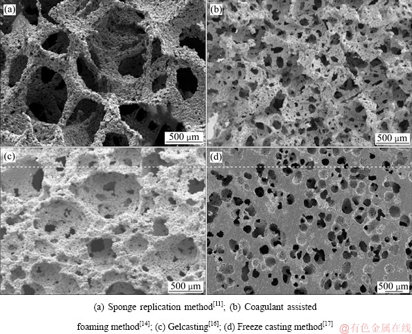
ASHBY��[5]����ĭ�������Ʊ����ɳ�4������(����ݷ���ˮ��Һ���������Һ̬����������̬������)������9�ֹ��շ��������У�ҽ�ö���Ѽ���Ͻ���Ʊ�������������ݷ����л����ิ�Ʒ������Ϸ��ݷ�������עģ�����䶳���취�������Ӹ��ºϳɷ����ŵ�������սᷨ��������������������취����ά�ϳɷ��ȡ����Ľ��Զ���ѵ��Ʊ����о���״���з���������������Ϊ�������Ĺ������ṩ �ο���
1 ����ѵ��Ʊ������������ص�
1.1 ����ɿ�
�����۵�ߡ����Դ����ó����������ݷ��Ʊ�����ѡ�OPPENHEIMER��[6]�������Ϊ�������壬��Ti6Al4V�Ͻ��ĩװ����������У�����պ��ٳ���0.33 MPa�������100 MPa�ȵȾ�ѹ(HIP)����840~1030 ����ѭ������ʹ����ڷ�ĩ�й����γɿ���ͬʱ��ĩ�������սᣬ�����γɿ�϶��8%~52%������ǿ��170~670 MPa������ģ��28~120 GPa����ĭ�ѺϽ�
�÷��Ʊ��Ķ���ѳɱ��������ڲ��ṹ�ֲ������ȣ����²�Ʒ���յĿ����ʽϵͣ���֮����ͨ���ڵijߴ��С�����м�ʮ�ף������ڹ���֯���ڲ����ij��룬����ò��ͼ1��ʾ����ˣ��÷������Ʊ�����ѷ�����о���Ӧ�ý���Ҳ��������������
ͼ1 ����ɿ��Ʊ�����ѵ�SEM��[6]
Fig. 1 SEM image of porous titanium prepared by gas phase pore formation [6]
1.2 ���ݼ��ɿ�
1.2.1 �л����ิ�Ʒ�
�л����ิ�Ʒ���ͨ���佬-����-����-�����ս�4�������Ʊ�����ѡ��Ƚ�TiH2��ĩ��ˮ�����������ȶ������������Ƴ�Һ�����ٽ��л���ĭ������Һ���г�ֽ�պ��ȡ�����ѹ��������Һ��������ȥ��ˮ�ݣ��������ս�����ͨ�����õ���ĭ�ѡ�
CACHINHO��[7]���ø÷����Ʊ����Ķ���ѿ�Ϊ100~600 ��m����϶��75%����ѹǿ�ȣ�23.72��1.12�� MPa������ģ��Ϊ(0.30��0.003) GPa�������ܽ�������(Sol-gel)��������Ʊ��ǻ���ʯ(HA)Ϳ�㣬��ģ����Һ�б��������õĿ�ʴ�ԡ�LEE��[8]������������(MAO)�ڶ���ѱ����Ʊ���HA/TiO2���Ϳ�㣬������ϸ������ʵ���������Ϳ��߱��Ϻõ�������ԡ����µ�[9]Ҳ�������������ױ�����
TANGE��[10]�Ըù��ս��иĽ������˽����ԵĿ���ò����ͨ�Լ���϶�ʣ����俹ѹǿ���뵯��ģ��ֻ��1.84~7.97 MPa��0.15~0.55 GPa����ѧ���ܽϲWANG��[11]���еĹ��ոĽ��ǽ����ѷ������ϩ��(PVA)���Һ�����������ս����ö���ѵĿ�ѹǿ���нϴ���ߣ�ԼΪ8.9~83.6 MPa��LIU��[12]���÷��Ʊ���Nb-Ti-Ta��Ͻ�ֲ���������ڣ���������ʾ����ֲ�����е�100~600 ��m��ͨ����ȫ�����¹���֯�ij��룬����֯ϸ���ڶ�Ͻ���������õ�ճ���������������Ҹ���ֲ���ϵ�����ǿ����ģ��(��ѹǿ��17.45~121.67 MPa������ģ��0.11~2.08 GPa)������ǵ���ѧƥ����Ҳ�������á�
�л����ิ�Ʒ��Ʊ��Ķ���ѣ���ͨ�����϶�ʶ��Ƚϸ�(��ͼ2(a))������ṹ����С�ͷֲ�ȡ���ں���ṹ���л���ĭ�Ǽ��ڽ��սα�������ȫ�����������»ӷ�����ɿױ��ڲ��пգ�ͬʱ�ڿױ����γ�6~45 ��m���ף����¿ױڵ���Ч��Ⱥ�ǿ���½�������Ӱ����ĭ��Ʒ����ѧ����[9]��
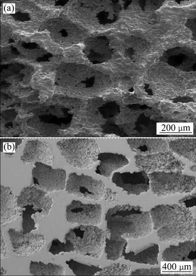
1.2.2 ���Ϸ��ݷ�
���Ϸ��ݷ��ǽ�������ĩ���ȶ��������ݼ���ˮ����Ƴɽ��ϣ��ڳ�ʪ�����³�ַ��ݣ�Ȼ����¸�������ս�õ�����ѡ�
KATO��[13]���ø÷��Ƶÿ�϶����17%~80%����ѹǿ���뵯��ģ���ֱ�Ϊ150~240 MPa��11~12 GPa�Ķ���ѡ�KAPAT��[14]���ü����������ݼ���һˮ�����������ȶ�������Ti6Al4V��ĩ����Ƴɽ��ϣ������¸�����1400 ���ս����ö���ѵĿ�϶�ʽ�ǰ���кܴ�������ԼΪ89.3%~65.2%������ΧԼΪ44.6~653.8 ��m����ѹǿ���뵯��ģ���ֱ���2.46~65.5 MPa��7.3~98.5 GPa��
�÷����ö������Ʒ�Ŀ�϶��ͨ�����Ƚϸߣ�������ͨ���ڵijߴ�Ҳ�Ƚϴ���֯�ܹ����룬�������γɵİ�����С���ֲ��ľ����Ժ��ѿ���(��ͼ2(b))�����ҽ����е��л��������C��OԪ�����ھ��縻������ռ�ȿɴ�2.38%~2.49%��1.37%~1.49%������ĭ��Ʒ����ѧ����Ӱ��ϴ�[14]��
1.2.3 ����עģ��
����עģ��������������������ʵ���ҿ�����һ���Ʊ��մɵĹ��շ������÷��Ʊ�������ǽ��л����塢��������ȥ����ˮ��Ti��ĩ����Ƴ�Һ���������������������������ٽ��裬Ȼ���Һע��ģ�����ܷ⣬�������½���ʹ���Һ��ַ��ݺ�ע���ͣ�������ս�õ���ĭ�ѡ�
ERK��[15]���ô˷��Ʊ�����ĭ�ѿ�϶��Ϊ4%~44%������ǿ����߿ɴ�200 MPa��SINGH��[4]�ø÷��Ƶö��������϶����������ߵ�72%~88%�����ֲ�Ϊ269~688 ��m������϶�ʵ����ȴ��������ǿ���뵯��ģ�������½���BIASETTO��[16]�õ�����Ϊ���ݼ�������ά��ˮ��Һ���ȶ�����Ti6Al4V��ĩ�佬��80 ���µ����еİ��������ȷ��ݲ��������پ�������ս�õ��Ķ���ѣ���϶�ʼ����ֲ�����ǰ���������ѹǿ�ȱ�����24.4~79.1 MPa��
�÷��ɶԲ�Ʒ�ṹ������ƣ����ò�Ʒ�Ŀ����ʺܸ�����ͨ�����ã������Ʊ������л���ԭ���к����л������������Ԫ�أ�����TiyOx��TiC�ȸ���������նԲ��ϵ�ʹ��ǿ�ȼ�ģ�����Σ������һ������ǰ�����Ϸ��ݷ����ڵ��������ƣ�����ò��ͼ2(c)��
ͼ2 ���ݷ��Ʊ��Ķ����SEM��
Fig. 2 SEM images of porous titanium prepared by foaming method
1.2.4 �䶳���취
�䶳���취�ǽ��䶳��(����ˮ����ϩ)����ɢ�����ϼ����ѷ�ĩ���л����ĥ������ո������н�������ֱ������ĥ�β�����������ȫ�ѳ���Ȼ��ע��ģ�����䶳ʹ�䶳����ֶ�������ģ������ս���͡�
JUNG��[17-18]����ϩ��Ϊ�䶳�����;۾�����Ϊ��ɢ�������ѷ�ĩ��ϣ���55 ������ĥ30 min��ע�����Ƶ�����Բ����ģ���У���ע����44 ��ģ���ڱ���30 r/minת������ɣ�֮��ģ����������ת��ֹ�����������ڼ���ϩ�����κ˳���ʹ���Ϲ̻����ͣ���ģ����200 MPa����Ⱦ�ѹʹ�������ȫ���ܻ����䶳������پ�1300 ������ս�2 h����ÿ�϶��Ϊ52%~71%����Ϊ95~362 ��m����ѹǿ�Ⱥ͵���ģ���ֱ�Ϊ57~183 MPa��1.3~5.0 GPa�Ķ���ѡ�WANG��[19]���ù����Ʊ��Ķ���Ѿ�����������ֲ���ùɹǽ��л���ʵ�飬֤���������������������Բ������ڹ���֯����ֲ�������л��Էֻ��������γ���ֲ��-����ǵ�Ƕ���塣
�䶳���취��Ԥ����Ʋ�Ʒ�ijߴ�����״�������ڴ�����λ����ֲ��֮���ƥ�䣻������ϩ�����ڶ��ɽα��Ƴ������������ṹ��ǿ���½�������̮��[17]����ͼ2(d)��ʾ���䶳���취���Ʊ������ߴ�Ͼ��ȵIJ�Ʒ���������ķֲ������Խϲ���²�Ʒ�Ŀ����ʽ��͡����⣬��ϩ�������ж��ԣ�����˿������䶳����չ�����о���
1.3 ����ɿ�
1.3.1 �ŵ�������սᷨ
�ŵ�������ս�(SPS)��һ�ַ�ĩұ���սἼ�����÷����Ʊ�������ǽ��ѷ���������ϼ����Ȼ�Ϻ�װ��ʯīģ���У��ս�ʱģ������ʩ�������ѹ��ģ���ڵķ�ĩ���˷ŵ������������ʹ��ĩ�������£��ս�ͬʱʩ��һ��ѹ��ʹ�������ܣ���һ�ָ�Ч�ķ�ĩ�ս��Ʊ�������
QUAN��[20]��Ti6Al4V��ĩ���������ϩ�����װ��Բ����ʯīģ�ߣ�700 ���¼�ѹ50 MPa�ս�8 min�õ���ʵ���壬��ˮ���ܽ�ȥ��NaCl������پ������ѹ�ս����õĶ�Ͻ��϶��Ϊ44.7%~70.0%�����ֲ�125~250 ��m������ǿ���뵯��ģ���ֱ�Ϊ43.0~110.2 MPa��9.5~33.0 GPa������ϸ������ʵ����ʾ���ϸ������������YAMANOGLU��[21]����ֱ�ӽ�Ti5Al2.5Fe�Ͻ����ѹ�ս�õ���϶��Ϊ28.4%~29.1%�Ķ�Ͻ��俹ѹǿ����ߴ�300 MPa����������Ʊ���HAͿ�㾭����֤�߱����õ����������ԡ�
SPS�ڷ�ĩ�ս᷽���������¿졢Ч�ʸߵľ����ƣ�ͨ�������Ԥ�ȿ��ƿ���С���������˿�϶�ʣ��ҿ�֮�����ͨ�Խϴ�ߴ細��Ϊ��[20](��ͼ3(a))�������úϽ���ֱ�Ӽ�ѹ�ս���ͻ�ö�ײ��ϣ������϶�ʹ��ͣ���������֯�ij��롣
1.3.2 ���������
����������ǽ�������ĩ�������Ϻ�ѹ�����ͻ����ʵ�������壬ȥ�����������ս���͡��÷������ս�Ϊ������ͱ��о���Ӧ���ڶ���ѵ��Ʊ��ϡ�
WEN��[22]��200~600 ��m̼�����������봿�ѷ۾��Ȼ�ϣ�100 MPa��ѹ���ͣ���200 ���±���5 hʹ����ӷ���Ȼ����1200 ���ս�2 h��ÿ�϶��Ϊ78%����ѹǿ���뵯��ģ���ֱ���35 MPa��5.3 GPa�Ķ�ײ��ϣ�IMWINKELRIED[23]��̼����刺�����60%~65%��϶�ʶ����Ӧ�õ����༹��ֲ����Ʊ��ϣ����ò�Ʒ�Ŀ�ѹǿ����߿ɴﵽ200 MPa��HSU��[24-25]������ͬ�ķ����Ƶ�Ti-7.5Mo��Ͻ𣬲��Ա���������������˾����������������Ե������ʯͿ�㡣LUPPO��[26]��̼�����������Ʊ���Zr-Ti-Nb��Ͻ���б�����Ժ��������ֹ���ֲʵ�飬�������֯����ֲ��ij��ϱ������á�DUNAND��[27]��NaClΪ����봿�ѷۻ���Ƶÿ�϶��42%~51%�Ķ���ѣ�����ģ����29~39 GPa��LI��[28]���������ѱ������������Ϳ����Ʊ���������ϸ������ʵ��֤ʵ������Ϳ��������õ���������������ԡ�PRADO��[29]�����ؿ�������Ѳ����б������������ϸ������ʵ������ò��������õ�������ԡ����������������������������ѵ��о�[30]�������ý���������롣
������Ʊ����㣬����ò�����ߴ缰��϶�ʶ���ͨ������ijߴ硢��״�����������е��ڣ��Ʊ��������ڿ��ƣ��ɻ���������ܵĶ���ѣ��ǵ�ǰ������Ʊ�Ӧ�÷dz��㷺��һ�ַ�������Ʒ��ò��ͼ3(b)�����÷������Ʊ��ϴ�ߴ缰������״��Ʒʱ��ռ���ƣ����ҽ�����봿�ѷ�ֱ�ӻ���ƵõĶ�ײ��ϣ��տ����������[27]����ˣ������߿�϶�Ų�����������Լ���Ч���پֲ�����տף�Ҳ�Ǹ÷�����Ҫ�����Ҫ���⡣
ͼ3 SPS�ս�Ti6Al4V��ĭ�Ͻ�SEM��[20]��������Ʊ�����ĭ��SEM��[27]
Fig. 3 SEM images of Ti6Al4V foams of the spark plasma sintered[20](a) and titanium foams created space holder method[27](b)
1.4 ���ʳɿ�
1.4.1 �������취
3D��ӡ�������취������ѡ���Լ����ս�(SLS)��ѡ���Լ����ۻ�(SLM)������̻�������(LENS)��������ѡ���ۻ�(EBM)��3D��ά�����Ȳ�ͬ����;������Ҫ����ΪCADģ����ơ�Ƭ����ɢ����������ӡ������������SLS��SLM��EBM�����ڶ���ѺϽ���Ʊ���Ӧ����ࡣ
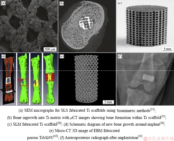
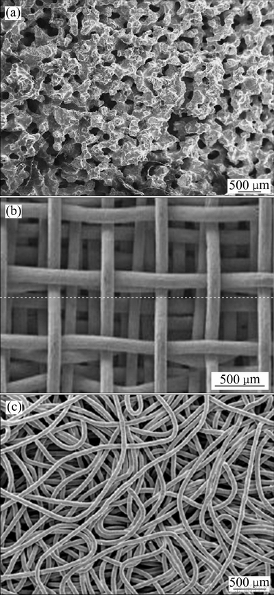
ѡ���Լ����ս�(SLS)���Լ�����Ϊ����Դ��ͨ��Ԥ�����ģ�ͺͳ�����ƶԹ����ĩ������ѡ���Ե�����ս���ͣ����ڹ��ʽ�С�����ò�Ʒ������[31]��SHISHKOVSKY[32]���������SLS�������Ƴ�Ӧ���ڿ�ǻ������Ķ���ѺϽ𡣶�SLS�Ʊ��Ŀ�϶��72%�Ķ���Ѿ��������HAͿ���[33]����ɽ��ǽ���ֲ��ʵ���еı��ֳ���������Ľ��(��ͼ4(a)��(b))��
ѡ���Լ����ۻ�����(SLM)��ѡ���Լ����սἼ��(SLS)ԭ�����ƣ���SLM���ʴ�Ч�ʸߣ���Ʒ������SLSҲ�������[31]��AMIN��[34]��SLM�Ʊ���Ti6Al4V��Ͻ����϶��Ϊ68%~84%�����ֲ�480~600 ��m[35]���������������ڲ��ϱ�����ȡ��TiO2���Բ㣬Ҳ�ѱ��ܶ��о���Ա֤ʵ���кܺõ�������ԣ�ϸ������������ֳ�ֻ�Ч�����������JAN��[36]��SLM�Ʊ�����״Ti6Al4V��Ͻ�����ɽ���ȹ�ȱ����ֲ��ʵ����(��ͼ4(c)��(d))����������ʾ��ֲ�������֯�������ã���������ʱ��Խ����ɽ���ȹ�����Ƕ�����ģ����ǿ�Ȼ����������͡�PEI��[37]�ڹ��ɹ���ֲʵ����Ҳ֤ʵ�˹�ϸ���ڸö����ֲ���Ͼ��кܺõ���ֳ�ֻ�Ч����
ͼ4 3D��ӡ����ѽṹ������ֲӦ��
Fig. 4 3D printing porous titanium and its transplantation application
������ѡ���ۻ�(EBM)�����ø������ܶȼ��������ʵĵ��������ѷ�ĩ����ɨ���ۻ��������������ά����Ѳ���[38]��MURR��[39]��EBM�����Ʊ�����϶��55%~89%�Ķ���ѺϽ�KNORRA��[40]��EBM�Ʊ���Ti6Al4V��Ͻ�ͨ����ѧ�������(CVD)�������ɹ��Ʊ��˾��ȵ�̼����Ϳ�㣬�һ�����Ͼ��ȳ�������ѧ���ܲ�δ���͡������ֶ�Ͻ�[41]����ɽ�������ֲʵ��(��ͼ4(e)��(f))�����������¹���֯���γɼ������϶��кܴ���ߣ�ͨ��������ͬ������Բ�ɽ�һ������ϸ������ֲ���ϱ��渽�š���ֳ���ֻ����Ӷ�����ϸ��������[42-43]��HARA��[44]��EBM�Ʊ��IJ�ͬ����Ͻ����ùɹ���ֲʵ������֤��800 ��m���Ķ��ֲ����Թ���֯�ij������źܺõĴٽ����á�
�������켼�����Ʊ����ӽṹ������м�������ƣ��ɶԹ�ȱ�ݻ��ߵ�ʵ������λ���и��Ի���ƣ���������״��ߴ�ֲ������ռ�ֲ���ʽ����϶�ʣ��ױں�ȵȣ�ʹ��Ʒ���õ��������λ����ƥ�䡣SLS��SLM�Ʊ��������ƣ����߱�ǰ�߹��ʸ���Ч�ʸ��ߣ����������Ʊ���������ʹC��H��OԪ���ھ��紦ƫ�ۣ����Ʊ���Ʒ�Ŀױ������ײ�����϶�������˿ױڵ���Ч��Ⱥ���ѧ����[31]�������������Ʊ�����Ѷ��ԣ��豸�����ϼ����չ淶����Ҫ��һ���о��ͷ�չ[45-46]��
1.4.2 ��ά�ϳɷ�
��ά�ϳɷ��ǽ���˿�������γ�������״���پ��������ս�õ���״����ѡ��ع���[47]��ֱ��Ϊ0.08~0.15 mm�Ĵ���˿�����������״����20~120 MPaѹ�����ͣ�1200 ���ս�õ���϶�ֲ�50~200 ��m����϶��Ϊ48%~82%������ǿ��12.6~180.4 MPa������ģ��0.33~1.05 GPa�Ķ�����������Ըýṹע������������ݵ������Ϳ��������ù�ػ����Һ���ϳ�����-����ѻ���壬�ϳɲ��ϵ�ҩ���ͷ�������ģ����Һ�б�������[48]��LI��[49]������ά��850�澭�����ɢ�Ƴ���״����ṹ�Ķ���ѣ����϶��Ϊ30%~ 70%����100~650 ��m������ģ��������ǿ�ȷֱ�Ϊ1.5~7.0 GPa��10~110 MPa���ùǹؽڵ���ֲ��ʵ�鱻֤�����ֲ��Ͼ������õ�ȱ�ݹ�����������[50]��
��ά�ս��Ʊ��Ķ����ǿ�������ҵ���ģ���ϵͣ��Ʊ����̼��ɱ��ͣ��������սᷨ�õ��IJ�����������(��ͼ5(c))���ڲ���ά�������ң�����ṹ��Ϊ��ɢ��������ģ��ƫ�ͣ������������ϲ�[47]��ͼ5(b)��ʾΪ100~200 ��mֱ������ά���й����֯���پ������ɢ���ӻ�õijߴ��ȶ�����ͨ�����ã�����ṹΪȫ������ά��״����ѣ��Ҹ������ܶ�����ά�����ս�Ķ����������ߡ������ڳ��ع������� ����ѧ������Ȼ��������������˽϶�Ӧ���ڶ�ǿ�ȼ�����ģ��Ҫ��ϵ͵�����λ�������ۿ�������
ͼ5 SHS�Ʊ���Ni-Ti��ĭ�Ͻ�[51]����ά��֯�Ʊ��Ķ����״[50]����ά�����Ʊ������״��SEM��[47]
Fig. 5 SEM images of nickel-titanium foam prepared by SHS[51](a), mesh porous titanium prepared by fiber weaving[50](b) and mesh porous titanium prepared by fiber tangled[47](c)
1.4.3 �����Ӹ��ºϳɷ�
�����Ӹ��ºϳɷ�(SHS)��������Ӧϵͳ�ڵ���ά�ַ�Ӧ�Ʊ���ײ��ϡ�ARCINIEGAS��[51]�ø÷��Ʊ�����϶��65%~70%��NiTiҽ����ĭ�Ͻ𣬿��ֲ�Ϊ370~440 ��m���������ǿ�ȿɴ�(142.5��29.3) MPa������ģ��(1.21��0.31) GPa��ƣ��ѭ�������ɴ�1��108��ARAKAWA��[52]��Al��Ti��Ϊ����Ԫ�أ�B4C�����ȼ�����400 ����ȼ���ϳɵ�Al-Ti��ĭ�Ͻ��϶��Ϊ60%~70%��
�����ӷ��Ʊ�����ĭ�Ͻ���нϸ߿�϶�ʺͽϺ���ͨ�ԣ���ѧ����Ҳ������ֲ��������ò��ͼ5(a)��ʾ��������ȼ�ղ�����˲����¿�ʹ������ĩ�ۻ������ײ���ƫ������ȱ�ݣ��Ʊ����̽��ѿ��ƣ���ˣ��÷����Ʊ�ҽ����ĭ�ѺϽ��汨�����١�
1.5 ����������Ʊ������
��Զ���ѿ�϶��״�����ֲ������������γɱտ����⣬�����������øĽ���������Ʊ��˿�϶�ֲ����ȡ���״����Ķ����ѡ�
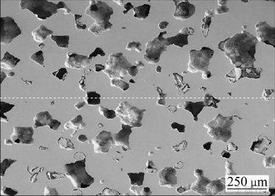
��NaClΪԭ�ϣ����ۺ�����ˮ�����Ϊճ�ϼ���ͨ����ԡ���Ⱥͻ�е�����Ʊ�NaCl���裬��740 ���ս��������״�������ͼ6��ʾ���������ս��NaCl��֮�����ʮ�����ף���Щ������������������п����ܽ�ȥ����
ɸѡ1000~2360 ��m��������Ϊ����������ĩ����װ��Ԥ��ģ�ߣ�����AUTOTAP��ʵ�ܶ��ǽ��ѷ�����䵽Ԥ������õġ��δ�����϶��(���չ��̼�ͼ7)��ͨ����ĩ�½��ĸ߶��ж�����Ƿ���ɡ�����ᆳ��ѹ���ͻ�á����-��ĩ�������壬������ˮ�ܽ�ȥ������õ���ά��ͨ�Ķ���������壬��1400 ���ս�2 h��ÿ����ȿɿصĶ���ѡ�
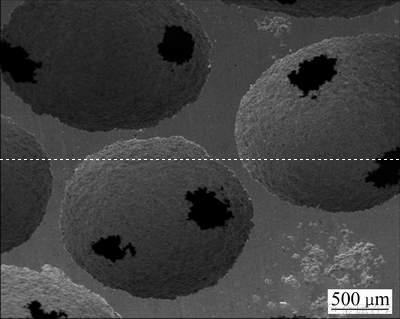
�����ֱ�ӻ��Ϸ����ö�ײ��ϣ��Ľ������Ʊ��Ķ���Ѱ���������Ԥ�����������������֮��ƽ����4~6����ͨ���ڣ�����ܽ�ȥ�����ף��տ��١��ҽṹ���ȿɿء������Ժã����ö���ѵĿ�϶��Ϊ70%~80%(��ͼ8)��
������Ʊ����������ܡ��������ڱ�1�����У�����������ݷ���SHS����SPS�����ڹ��ձ������ɱ�ԭ���½������������١����Ϸ��ݷ�������עģ�������о����ڳ��ԸĽ������ิ�Ʒ����䶳���취����������������취����ά�ϳɷ��ȹ����Կ���������һֱ�����о���λ������ǰ�ƽ������ֹ��ղ����ѽ������ʵ����ٴ�Ӧ�ýΡ�

ͼ6 �ս��NaCl�����SEM��
Fig. 6 SEM images of sintered NaCl Beads
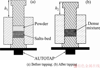
ͼ7 ��ṹ��������ʾ��ͼ
Fig. 7 Schematic process of porous structure construction
ͼ8 �ս�����ѵ�SEM��
Fig. 8 SEM image of sintered porous titanium
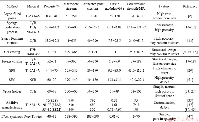
��1 ����Ѽ���Ͻ��Ʊ����������ܸ�Ҫ
Table 1 Summary of preparation process and properties of porous titanium and its alloys
2 ����ѵ�Ӧ�øſ�
��ǰ��������������ٴ���������ǣ��ǣ�ϥ�ؽڣ��Źؽڵ����ܲ�������˵Ĺ���֯��λ��
PENG��[53]���þ���ϩͿ����ԵĶ���������϶Ի�е�Դ��˵����ۿ����۵Ļ��߽��������ƣ�����Ի��ߵĻָ�������ټ�⣬�����������������϶��ۿ����ؽ����г��ڵĻ�е�ȶ��ԣ��ܹ����������ݣ����ӣ���������֢�ȷ��ս�����͡�
WINTHER��[54]���ø߿�϶�ʶ���ѶԻ���ʵʩȫϥ�ؽ��û���(TKA)�������ڶ���ֲ����λ��λ��������м�⣬�����ʾ�������Ǩ�����ʷdz��ͣ��³���Ҳ�dz�С��HARWIN��[55]Ҳ��219�����ߵ�ȫϥ�ؽ��û�(TKA)������Ҳ֤ʵ�����ָ߿�϶����ѵĻ�е�ȶ�������������QASSEMYAR��[56]����3D��ӡ�Ķ�������Ǽ�����������Ļ��߽������ؽ�������ʹ�����沿�������Լ����ָܻ�������������һ��ʱ�ڵĻ����������������̺ۣ��������ּ����кܺõ���֯����������������������ϱ��ֳ����õ�Ӧ��ǰ����
DELANOIS��[57]���ø߿�϶����Ѳ��϶�35�����仼�ߵ��Źؽ�ʵʩ�û�����������5�����ϵ��ټ�⣬����һ�����߳��ֲ�����Ⱦ���������߳��ְ�Ѫ��������ָ�������������ã�֤�����ֶ���Ѿʱ����Źؽھ��кܺõ������������ڵı�������ҽ�ƹ�˾��2016���Ƴ�3D��ӡ�Ķ�����˹���ͼ����ں���(��еע20163460859)�����ʳҩ�����֤���ɹ����뼹���û�ֲ�����˹������г����ò�Ʒӵ�����õ����������Ժ����������������ʹǵĵ���ģ���и��õ���ѧƥ���ԣ�Ŀǰʹ��������Ϊ�ȶ���
3 ����
�������Ϊ�������ֲ���ϣ���������������������ԣ������������滻���˵Ĺ���֯�����������з����������������ֲ����������ԣ�����ϸ���ĸ��š���ֳ�ͷֻ��������ӿ���֯�ڶ�ṹ�е��������ʣ�ʹ��ֲ����ԭ����֯���õس��ϣ��Դﵽ��ȱ�ݵ�Ŀ�ġ�
��ǰ���ڶ�������ֲʵ����ٴ�Ӧ���о��϶����Ҫ��3D��ӡ������������䶳���취����ά�ս�ȷ����Ʊ��Ķ���ѡ���ͬ�Ʊ��������и����ŵ㣬���ձ�����Ʊ��ɱ��ϸ�����ƫ�͵����⣻���⣬��Բ�ͬ���ߵ�ʵ�ʹ��������������Ҫ��ͬ��ѧ���ܼ���϶�ṹ�Ĺ���ֲ���ϣ���ˣ��ڽϴ�Χ�ڶԶ���ѹ���ֲ���Ͻṹ�͵���ģ�����е�����Ȼ������о����Ը��õؽ����ֲ���������λ����ѧƥ�����⡣
������ٴ�Ӧ����Ȼ����һ��ʱ�ڣ������������������ϵ��;��ԡ������ԡ���������Ȼ�д������ij��ڼ��顣
REFERENCES
[1] ��ϲ��. ��ֲ����ϵ�Ӧ�ü���չǰ��[J]. �й�ҽҩ�������������, 2007, 1(5): 30-33.
LIU Xi-chun. Application and development prospect of bone implant materials[J]. Chinese Medical Technology Economy and Management, 2007, 1(5): 30-33.
[2] LEWIS G. Properties of open-cell porous metals and alloys for orthopaedic applications[J]. Journal of Materials Science Materials in Medicine, 2013, 24 (10): 2293-2325.
[3] AGUILAR MAYA A E, GRANA D R, HAZARABEDIAN A, KOKUBU G A, LUPPO M I, VIGNA G. Zr-Ti-Nb porous alloys for biomedical application[J]. Materials Science & Engineering C, 2012, 32(2): 321-329.
[4] SINGH R, LEE P D, JONES J R, POOLOGASUNDARAMPILLAI G, POST T, LINDLEY T C, DASHWOOD R J. Hierarchically structured titanium foams for tissue scaffold applications[J]. Acta Biomaterialia, 2010, 6(12): 4596-4604.
[5] ASHBY M F, EVANS A G, FLECK N A, GIBSON L J, HUTCHINSON J W, WADLEY H N G. Metal foams: A design guide[J]. Applied Mechanics Reviews, 2001, 23(6): 119.
[6] OPPENHEIMER S, DUNAND D C. Solid-state foaming of Ti-6Al-4V by creep or superplastic expansion of argon-filled pores[J]. Acta Materialia, 2010, 58 (13): 4387-4397.
[7] CACHINHO S C, CORREIA R N. Titanium scaffolds for osteointegration: Mechanical, in vitro and corrosion behaviour[J]. Journal of Materials Science Materials in Medicine, 2008, 19(1): 451-457.
[8] LEE J H, KIM H E, KOH Y H. Highly porous titanium (Ti) scaffolds with bioactive microporous hydroxyapatite/TiO2, hybrid coating layer[J]. Materials Letters, 2009, 63(23): 1995-1998.
[9] GAO Y, XU X X, YANG Z M. Novel TiC/Ti open cellular foams prepared by a modified sponge-coating method using high frequency induction heating process[J]. Journal of Materials Science & Technology, 2013, 29(4): 339-343.
[10] TANGE M, MANONUKUL A, SRIKUDVIEN P. The effects of organic template and thickening agent on structure and mechanical properties of titanium foam fabricated by replica impregnation method[J]. Materials Science & Engineering A, 2015, 641: 54- 61.
[11] WANG C L, CHEN H J, ZHU X D, XIAO Z W, ZHANG K, ZHANG X D. An improved polymeric sponge replication method for biomedical porous titanium scaffolds[J]. Materials Science & Engineering C, 2017, 70(2): 1192-1199.
[12] LIU J, RUAN J M, CHANG L, YANG H L, RUAN W. Porous Nb-Ti-Ta alloy scaffolds for bone tissue engineering: Fabrication, mechanical properties and in vitro/vivo biocompatibility[J]. Materials Science & Engineering C, 2017, 78: 503-512.
[13] KATO K, OCHIAI S, YAMAMOTO A, DAIGO Y, HONMA K, MATANO S, OMORI K. Novel multilayer Ti foam with cortical bone strength and cytocompatibility[J]. Acta Biomaterialia, 2013, 9(3): 5802-5809.
[14] KAPAT K, SRIVAS P K, DHARA S. Coagulant assisted foaming��A method for cellular Ti6Al4V: Influence of microstructure on mechanical properties[J]. Materials Science & Engineering A, 2017, 689: 63-71.
[15] ERK K A, DUNAND D C, SHULL K R. Titanium with controllable pore fractions by thermoreversible gelcasting of TiH2[J]. Acta Materialia, 2008, 56(18): 5147-5157.
[16] BIASETTO L, MORAES E G, COLOMBO P, BONOLLO F. Ovalbumin as foaming agent for Ti6Al4V foams produced by gelcasting[J]. Journal of Alloys & Compounds, 2016, 687: 839-844.
[17] JUNG H D, YOOK S W, JANG T S, LI Y L, KIM H E, KOH Y H. Dynamic freeze casting for the production of porous titanium (Ti) scaffolds[J]. Materials Science & Engineering C, 2013, 33(1): 59-63.
[18] LEE H, JANG T S, SONG J, KIM H E, JUNG H D. Multi- scale porous Ti6Al4V scaffolds with enhanced strength and biocompatibility formed via dynamic freeze-casting coupled with micro-arc oxidation[J]. Materials Letters, 2016, 185: 21-24.
[19] WANG G H, FU H, ZHAO Y Z, ZHOU K C, ZHU S H. Bone integration properties of antibacterial biomimetic porous titanium implants[J]. Transactions of Nonferrous Metals Society of China, 2017, 27(9): 2007-2014.
[20] QUAN Y J, ZHANG F M, REBL H, NEBE B, KE��LER O, BURKEL E. Ti6Al4V foams fabricated by spark plasma sintering with post-heat treatment[J]. Materials Science & Engineering A, 2013, 565 (10):118-125.
[21] YAMANOGLU R, GULSOY N, OLEVSKY E A, GULSOY H O. Production of porous Ti5Al2.5Fe alloy via pressure-less spark plasma sintering[J]. Journal of Alloys & Compounds, 2016, 680: 654-658.
[22] WEN C E,MABUCHI M, YAMADA Y, SHIMOJIMA K, CHINO Y, ASAHINA T. Processing of biocompatible porous Ti and Mg[J]. Scripta Materialia, 2001, 45(10): 1147-1153.
[23] IMWINKELRIED T. Mechanical properties of open-pore titanium foam[J]. Journal of Biomedical Materials Research Part A, 2007, 81(4): 964-970.
[24] HSU H C, WU S C, HSU S K, TSAI M S, CHANG T Y, HO W F. Processing and mechanical properties of porous Ti-7.5Mo alloy[J]. Materials & Design, 2013, 47(9): 21-26.
[25] HSU H C, WU S C, HSU S K, CHANG T Y, HO W F. Effect of ball milling on properties of porous Ti-7.5Mo alloy for biomedical applications[J]. Journal of Alloys & Compounds, 2014, 582(2): 793-801.
[26] AGUILAR MAYA A E, GRANAD R, HAZARABEDIAN A, KOKUBU G A, LUPPO M I, VIGNA G. Zr-Ti-Nb porous alloys for biomedical application[J]. Materials Science & Engineering C, 2012, 32(2): 321-329.
[27] YE B, DUNAND D C. Titanium foams produced by solid-state replication of NaCl powders[J]. Materials Science & Engineering A, 2010, 528(2): 691-697.
[28] LI M T, WANG Y, GAO L L, SUN Y H, WANG J X, QU S X, DUAN K, WENG J, FENG B. Porous titanium scaffold surfaces modified with silver loaded gelatin microspheres and their antibacterial behavior[J]. Surface & Coatings Technology, 2016, 286: 140-147.
[29] PRADO R F, OLIVEIRA F S, NASCIMENTO R D, VASCONCELLOS L M R, CARVALHO Y R, CAIRO C A A. Osteoblast response to porous titanium and biomimetic surface: In vitro analysis[J]. Materials Science & Engineering C, 2015, 52: 194-203.
[30] CHEN Y H, KENT D, BERMINGHAM M, MANSHADI A D, DARGUSCH M. Manufacturing of biocompatible porous titanium scaffolds using a novel spherical sugar pellet space holder[J]. Materials Letters, 2017, 195(92): 92-95.
[31] ϼ, ��ѩ��, ������, ����Ƽ, ���²�, ��ѡ��. ����ҽ�ö���Ѽ��ѺϽ���ٳ����о���չ[J]. ���ϵ���, 2016, 30(7): 109-114.
XIE Fang-xia, HE Xue-ming, L�� Yan-ming, WU Mei-ping, HE Xin-bo, QU Xuan-hui. Research progress in laser rapid forming of porous titanium and its alloys for biomedical applications[J]. Materials Review, 2016, 30(7): 109-114.
[32]
[33] TAMADDON M, SAMIZADEH S, WANG L, BLUNN G, LIU C Z. Intrinsic osteoinductivity of porous titanium scaffold for bone tissue engineering[J]. International Journal of Biomaterials, 2017, 2017: 1-11.
[34] AMIN YAVARI S, CHAI Y C, BOTTGER A J, WAUTHLE R, SCHROOTEN J, WEINANS H, ZADPOOR A A. Effects of anodizing parameters and heat treatment on nano-topographical features, bioactivity, and cell culture response of additively manufactured porous titanium[J]. Materials Science & Engineering C, 2015, 51: 132-138.
[35] HEDAYATI R, AMIN Y S, ZADPOOR A A. Fatigue crack propagation in additively manufactured porous biomaterials[J]. Materials Science & Engineering C, 2017, 76: 457-463.
[36] JAN W D, LINDNER T, BERGSCHMIDT P, BADER R. Bio-mechanical stability of novel mechanically adapted open-porous titanium scaffolds in metatarsal bone defects of sheep[J]. Biomaterials, 2015, 46: 35-47.
[37] PEI X, ZHANG B Q, FAN Y G, ZHU X D, SUN Y, WANG Q G, ZHANG X D, ZHOU C C. Bionic mechanical design of titanium bone tissue implants and 3D printing manufacture[J]. Materials Letters, 2017, 208(128): 133-137.
[38] CANSIZOGLU O, HARRYSSON O, CORMIER D, WEST H, MAHALE T. Properties of Ti-6Al-4V non-stochastic lattice structures fabricated via electron beam melting[J]. Materials Science & Engineering A, 2008, 492(1/2): 468-474.
[39] MURR L E, GAYTAN S M, MEDINA F, MARTINEZ E, MARTINEZ J L, HERNANDEZ D H, MACHADO B I, RAMIREZ D A, WICKER R B. Characterization of Ti-6Al-4V open cellular foams fabricated by additive manufacturing using electron beam melting[J]. Materials Science & Engineering A, 2010, 527(7/8): 1861-1868.
[40] KNORR T, HEINL P, SCHWERDTFEGER J, KORNER C, SINGER R F, ETZOLD B J M. Process specific catalyst supports Selective electron beam melted cellular metal structures coated with microporous carbon[J]. Chemical Engineering Journal, 2012, 181/182(1): 725-733.
[41] HUANG H, LAN P H, ZHANG Y Q, LI X K, ZHANG X, YUAN C F, ZHENG X B, GUO Z. Surface characterization and in vivo performance of plasma-sprayed hydroxylapatite coated porous Ti6Al4V implants generated by electron beam melting[J]. Surface &Coatings Technology, 2015, 283: 80-88.
[42] LI X, MA X Y, FENG Y F, MA Z S, WANG J, MA T C, QI W, LEI W, WANG L. Osseointegration of chitosan coated porous titanium alloy implant by reactive oxygen species-mediated activation of the PI3K/AKT pathway under diabetic conditions[J]. Biomaterials, 2015, 36: 44-54.
[43] WANG L, HU X F, MA X Y, MA Z S, ZHANG Y, LUA Y Z, LI X, LEI W, FENG Y F. Promotion of osteointegration under diabetic conditions by tantalum coating-based surface modification on 3-dimensional printed porous titanium implants[J]. Colloids & Surfaces B Biointerfaces, 2016, 148: 440-452.
[44] HARA D, NAKASHIMA Y, SATO Y, HIRATA M, KANAZAWA M, KOHNO Y, YOSHIMOTO K, YOSHIHARA Y, NAKAMURA A, NAKAO Y, IWAMOTO Y. Bone bonding strength of diamond-structured porous titanium alloy implants manufactured using the electron beam melting technique[J]. Materials Science & Engineering C, 2016, 59: 1047-1052.
[45] Ф ��, ���. ��ĭ�����ѵ��Ʊ������о���չ[J]. ϡ�н��������빤��, 2017(6): 1734-1748.
XIAO Jian, QIU Gui-bao. Research progress in preparation methods of titanium foams or porous titanium[J]. Rare Metal Materials and Engineering, 2017(6): 1734-1748.
[46] LIU Y J, LI S J, WANG H L, HOU W T, HAO Y L, YANG R, SERCOMBE T B, ZHANG L C. Microstructure, defects and mechanical behavior of beta-type titanium porous structures manufactured by electron beam melting and selective laser melting[J]. Acta Materialia, 2016, 113: 56-67.
[47] HE G, LIU P, TAN Q. Porous titanium materials with entangled wire structure for load-bearing biomedical applications[J]. Journal of the Mechanical Behavior of Biomedical Materials, 2012, 5(1): 16-31.
[48] LI Q, HE G. Gelatin-enhanced porous titanium loaded with gentamicin sulphate and in vitro release behavior[J]. Materials & Design, 2016, 99: 459-466.
[49] LI F P, LI J S, XU G S, LIU G J, KOU H C, ZHOU L. Fabrication, pore structure and compressive of behavior anisotropic porous titanium for human trabecular bone implant applications[J]. Journal of the Mechanical Behavior of Biomedical Materials, 2015, 46: 104-114.
[50] CHANG B, SONG W, HAN T X, YAN J, LI F P, ZHAO L Z, KOU H C, ZHANG Y M. Influence of pore size of porous titanium fabricated by vacuum diffusion bonding of titanium meshes on cell penetration and bone ingrowth[J]. Acta Biomaterialia, 2016, 33: 311-321.
[51] ARCINIEGAS M, APARICIO C, MANERO J M, GIL F J. Low elastic modulus metals for joint prosthesis: Tantalum and nickel-titanium foams[J]. Journal of the European Ceramic Society, 2007, 27(11): 3391-3398.
[52] ARAKAWA Y, KOBASHI M, KANETAKE N. Foaming behavior of long-scale Al-Ti intermetallic foam by SHS mode combustion reaction[J]. Intermetallics, 2013, 41(10): 22-27.
[53] PENG M Y, MERBS S L, GRANT M P, MAHONEY N R. Orbital fracture repair outcomes with preformed titanium mesh implants and comparison to porous polyethylene coated titanium sheets[J]. Journal of Cranio-maxillo-facial Surgery, 2016, 45(2): 271-274.
[54] WINTHER N S, JENSEN C L, JENSEN C M, LIND T, SCHRODER H M, FLIVIK G, PETERSEN M M. Comparison of a novel porous titanium construct (Regenerex ) to a well proven porous coated tibial surface in cementless total knee arthroplasty��A prospective randomized RSA study with two-year follow-up[J]. The Knee, 2016, 23(6): 1002-1011.
[55] HARWIN S F, PATEL N K, FRCS, CHUGHTAI M, KHLOPAS A, RAMKUMAR P N, ROCHE M, MONT M A. Outcomes of newer generation cementless total knee arthroplasty: Beaded periapatite-coated vs highly porous titanium-coated implants[J]. The Journal of Arthroplasty, 2017, 32: 2156-2160.
[56] QASSEMYAR Q, ASSOULY N, TEMAM S, KOLB F. Use of a three-dimensional custom-made porous titanium prosthesis for mandibular body reconstruction[J]. International Journal of Oral & Maxillofacial Surgery, 2017, 46(10): 1248-1251.
[57] DELANOIS R E, GWAM C U, MOHAMED N, KHLOPAS A, CHUGHTAI M, MALKANI A L, MONT M A. Midterm outcomes of revision total hip arthroplasty with the use of a multihole highly-porous titanium shell[J]. The Journal of Arthroplasty, 2017, 32: 2806-2809.
Preparation and development of porous titanium material for bone transplanting
JIA Jian-gang, JING Yong-zhi, GAO Chang-qi, LIU Chang, JI Gen-shun, GUO Tie-ming
(Lanzhou University of Technology, State Key Laboratory of Advanced Processing and Recycling of Non-ferrous Metals, Lanzhou 730050, China)
Abstract: The porous titanium is a porous metal material with a three-dimensional open interconnected pore structure. Compared with dense biometal materials, its elastic modulus is greatly reduced due to its openability and connectivity, and thus it can be well matched to human bones, at the same time, free circulation of body fluids, combined with good biocompatibility of titanium, thus the porous titanium is used as orthopedic transplant material. How to control the porous structure and obtain the porous titanium matrix with excellent performance is the premise and foundation for carry out related research and application. The porous titanium alloy have been developed, and the preparation methods conclude gas foaming method, organic sponge replication method, slurry foaming method, gel column molding method, frozen casting method, self-propagating high-temperature synthesis method, and spark plasma sintering method, added pore-forming agent method, additive manufacturing method, fiber synthesis method, etc. Different preparation methods have their own advantages, but most of them suffer problems, such as high preparation cost or low performance. The orthopedic transplant material with different mechanical properties and pore structure are required for the actual bone condition of different patients. Therefore, adjusting the structure and modulus of porous titanium for bone-transplanting in a large range still will be studied. The porous titanium bone graft materials have achieved clinical application, the porous titanium vertebral implant materials are rolled out by domestic institutions. Porous titanium as a bone graft material, there is still much room for research and clinical applications in the future.
Key words: porous titanium; foams metal; bone graft material; porous preparation; biocompatibility
Foundation item: Project(51461029) supported by the National Natural Science Foundation of China; Project (1602GKDD012) supported Major Special of Science & Technology of Gansu Province, China
Received date: 2018-06-12; Accepted date: 2019-03-06
Corresponding author: JIA Jian-gang; Tel: +86-13919809312; E-mail: lzhjiajiangang@163.com
(�༭ �� ��)
������Ŀ��������Ȼ��ѧ����������Ŀ(51461029)������ʡ�Ƽ��ش�ר��ƻ���Ŀ(1602GKDD012)
�ո����ڣ�2018-06-12�������ڣ�2019-03-06
ͨ�����ߣ��ֽ��գ������ڣ���ʿ���绰��13919809312��E-mail��lzhjiajiangang@163.com
ժ Ҫ���������һ�־�����ά��ͨ�ṹ�Ľ������ϣ��������������������ȣ����������Ŀ����Ժ���ͨ��ʹ�䵯��ģ������Ƚ��ͣ��Ӷ��ܹ��������ģ���Ϻ�ƥ�䣬ͬʱʹ��Һ������ͨ����֮�Ѿ������õ����������ԣ�����������ǿ���ֲ���ϡ�����ο��ƶ�ṹ�������������Ķ���ѻ����ǿ�չ����о���Ӧ�õ�ǰ����������ѿ������Ķ���Ѽ���Ͻ���Ʊ�������������ݷ����л����ิ�Ʒ������Ϸ��ݷ���������ģ�����䶳���취�������Ӹ��ºϳɷ����ŵ�������սᷨ��������������������취����ά�ϳɷ��ȡ���ͬ�Ʊ��������и����ŵ㣬���ձ�����Ʊ��ɱ��ϸ�����ƫ�͵����⣻���⣬��Բ�ͬ���ߵ�ʵ�ʹ����������Ҫ��ͬ��ѧ���ܼ���϶�ṹ�Ĺ���ֲ���ϡ���ˣ��ڽϴ�Χ�ڶԶ���ѹ���ֲ���Ͻṹ��ģ�����е�����Ȼ������о���Ŀǰ������ѹ���ֲ������ʵ���ٴ�Ӧ�ã����ڻ���Ҳ�Ѿ��Ƴ��������ֲ����������ٴ�ʵ����δ�����������Ϊ����ֲ���ϵ��о����ٴ�Ӧ�����кܴ�ռ䡣
