
DOI��10.19476/j.ysxb.1004.0609.2017.09.18
̼����ͭ�������ıȱ�����Ϳṹ
����1��κ��ǿ1, 2����ѧ��1��������1���� ��1, 2��������1
(1. ����������ѧ ��ѧԺ������ 730050��
2. ����������ѧ ʡ��������ɫ�����Ƚ��ӹ��������ù����ص�ʵ���ң����� 730050)
ժ Ҫ��
����ֱ���绡�ŵ�������弼���Ʊ�̼����ͭ��������������Ʒ����ò������ṹ�����ȡ��ȱ�����Ϳṹ���ø߷ֱ����������(HRTEM)��X����������(XRD)��N2��-�Ѹ��Ȳ����ֶν��з��������������ֱ���绡�������弼���Ʊ���̼����ͭ���������е��͵ĺ˿��ͽṹ���ں�Ϊ���������Ľ���ͭ�����Ϊʯī̼�㡣������Ҫ�����λ������Σ�������ԱȽϾ��ȣ���ɢ�����ã������ֲ���20~100 nm��Χ�ڣ�ƽ������Ϊ50 nm�����̼��ĺ��Ϊ10 nm��̼����ͭ�������ĵ����������������ͣ�����֮��Ŀ�϶�Խ��Ϊ������Ʒ��BET�ȱ����Ϊ33 m2/g����������Ϊ45 nm����TEM��XRD��õĽ������һ�¡�BJH�����ۻ��ܿ�����BJH����ƽ�����ֱ�Ϊ0.112 cm3/g��13 nm��
�ؼ��ʣ�
̼�������˿ǽṹ�����������ȱ�������ṹ��
���±�ţ�1004-0609(2017)-09-1896-06���� ��ͼ����ţ�TB383���� ���ױ�־�룺A
�������Ƕ����ײ����о��IJ������룬������ʶ���Ʊ����������������׳߶��Ͻ����˹���ơ��ɿغϳɾ��ж��ص���̬�ṹ�����ʵ������ϲ��Ϸ�����ɡ���������ͭ�۾������ӳߴ�ЧӦ��С�ߴ�ЧӦ������ЧӦ�ͺ����������ЧӦ�����ԣ����ֳ���ͬ�������Ӻͳ��������ϵĹ⡢�硢�ȡ��ŵ��������ʺͻ�ѧ����[1-5]������ͭ����Ϊһ�����͵Ĺ��ܲ��ϣ��ڸ����ܵ缫���ϡ�����ӡˢ�������Ӽ����������������罬�ϡ���������������ϡ����浼��Ϳ�㴦���������������Ź㷺��Ӧ��ǰ��[6-9]����ʵ��Ӧ����Ҫ������ͭ�۾��д��ȸߡ���ɢ�Ժá����Ⱦ��ȵ��ص㣬���������������С������ԭ����ռ�����࣬�����ܸߣ�����������ռ���δ���ͼ���������ӽṹ����ѧ��Ӧ���Ժ�ǿ�������ܵ���绷����Ӱ��������ž����������¿����ȶ��Ժͻ��Խ��ͣ����ͨ����Ҫ�Խ����������ı�����а�������[10-15]��̼����������������һ�־��к˿ǽṹ�����ͽ�ײ��ϣ���Ҫ�д�С�ӽ�����λ�ߴ��������������λ��С���ն��Լ��ɶ������Χ�ɵĿ������͵Ŀṹ[16-17]����ײ��ϲ������кܴ�ıȱ�����ͽ�ǿ�������������������ͨ�Ŀṹ�ܳ�Ϊ���ʽ�������������ߵ�λ���������Ĵ�Ч��[18]�����ײ��ϵĽṹ��������ѧ������Ӱ�������ܵ���Ҫԭ�ȱ�����Ϳṹ�ֲ��Ǻ������ֳ�������Ϻͽ�ײ��ϵĻ��ԡ����������ȶ������ܵ�һ����Ҫ����ָ��[19-21]���о���ײ��ϵıȱ�����Ϳṹ���ﻯ������������֮��Ĺ�ϵ����ʵ���ҽεķ����Ʊ�����Ӧ�þ�����Ҫ���塣
������������ֱ���绡�������弼�����ڶ������������³ɹ��Ʊ�̼����ͭ�������������ø߷ֱ����������(HRTEM)��X����������(XRD)��N2��-�Ѹ��Ȳ����ֶζ���������ò������ṹ�����ȡ��ȱ�����Ϳṹ�����������˱�����
1 ʵ��
1.1 ��Ʒ���Ʊ�
̼����ͭ�������������������Ƶ�ֱ���绡���������Ʊ�������������װ�ö����[22]��װ����Ҫ��ֱ����Դװ�á�����װ�á���շŵ��ҡ�����װ�á���ȴ�ռ�Ͳ����Ϊ�������������ˮ��ͭ����͵ȼ���������ɣ����Դ�ͳ�绡�ŵ緽�������˸Ľ������缫װ�ô�ˮƽ�ṹ��Ϊ��ֱ�ṹ����ʵ��Ϊ�˱��������¶ȹ����淢���ۻ����������ײ�������ˮ��ȴװ�ã�����ͭ�����ڲ����Ƕ��һ�������������õĴ�ʯī��������̼�ۺ�ͭ�۰���һ���ı�����Ϻ�Ϊԭ�Ϸ�����ʯī�����ڡ���ʯī��(ֱ��6 mm)���������ڷ�Ӧ���Ϸ��������缫��϶�������ķ���ͨ�뷴Ӧ������ʵ��ǰ���ȼ��װ����·����·��ˮ·������״̬�������缫�����ܷ�װ�á���������û�е�ú���ɢ�ó�����նȴ�1��10-3 Pa����β��øߴ�Ar(99.99%)��Ϊ���Թ����������ս��о�����ϴ����һ���������������������Ũ�ȡ�Ȼ��ͨ��Ar���Ա������գ�������Ӧ����ѹΪ1��102 Pa���缫����3~4 mm����ֱͨ����Դ����ȼ�绡�����Ƶ绡����120 A���绡��ѹ��25 V���ҡ������������缫֮��ĵ���������£�̼��ͭ������ڸ����±�Ѹ�ټ��������γ���������ɺܸߵľ�������������⽫���¾��ȵijɺ˹��̡��ڽӽ���ȴ�ռ�Ͳ�����У�����Ѹ����ʧ��������ȴ�����������γɺ˿ǽṹ��̼�������������������о�����������ƷΪ��Ar�����С���ѹ1 kPa���绡����120 A��ˮ��ȴ�������Ʊ���̼����ͭ��������
1.2 ��Ʒ�ı���
�����ձ���ѧRigaku��˾��D/Max-2400X������������(Cu��K������X���߲���Ϊ0.154056 nm��������ѹ40 kV������30 mA��ɨ�跶Χ20��~90�㣬ɨ���ٶ�12 (��)/min)��������������ṹ����������Scherrer��ʽ���㾧�����ȣ�����JEM-2010�߷ֱ����������(���ٵ�ѹ200 kV����ֱ���0.19 nm)�۲���Ʒ����ò����������ֲ�����������Micromerities��˾ASAP-2010�ͱ������������Dzⶨ��Ʒ�ıȱ�����Ϳṹ�����þ�̬���ݷ������ø���Ϊ0.0001 g��ƽȷ��ȡһ����������Ʒ������Ʒ���У�Ȼ���䰲װ�ڼ��ȶ��ϡ�����ն�С��1��10-3 Pa�������£���472~673 K�¶��£�����һ��ʱ��������������������������ԭ�е�������ٽ�������������ܷ������壬��N2Ϊ�����ʣ���Һ�������¶���(78 K)����Ʒ���о�̬�������������ѹ��p/p0��0.01~0.99֮������ĩ�ⲿ���ڲ�ͨ�ױ���Ե��������������ͬѹ���������������Ѹ������ɼ�������ƣ��ⶨ��Ʒ��N2�������Ѹ������ߡ�����BET����ģ�Ͳ��Թ�ʽ������Ʒ�ıȱ����������BJH����ģ�Ͳ�����Ʒ�Ŀ�����Ϳ��ֲ������� ������
2 ���������
2.1 ��Ʒ������
ͨ���Ա��������Ʊ���Ʒ����XRD�������о�̼����ͭ������������ṹ��ͼ1��ʾΪ��Ʒ��XRD�ס���ͼ1�п��Կ�������2��Ϊ43.29�㡢50.36�㡢74.02���90.04�㴦����4�����Ե���������塣��Щ��Ҫ������λ�ú�ǿ�������Ƭ(PDF No.04-0836)���������ṹͭ����������Ӧ���ֱ��Ӧ��ͭ��(111)��(200)��(220)��(311)���档˵����������Ҫ�ɷ���ͭ�����Ҿ������������ľ���ṹ����XRD���г���ͭ�����������⣬����2��Ϊ26.48�㴦�����������������壬��Ӧ����ʯī̼(111)���棬�����˷����Ʊ�����Ʒʯī���̶Ƚϸߡ�XRD������������Ʒ�г���̼�ͽ���ͭ�����������֮�⣬��û�й۲쵽����̼��������������Լ������������������Ĵ��ڣ���˵��̼���������������ӵ���Ҫ�ɷ���FCC�ṹ��ͭ��ʯī̼�����⣬����X�����������ۣ�ͨ�������İ�߿�����Scherrer��ʽd=0.89��/(Bcos��)���㱻�������������ṹͭ�˵ľ����ߴ磬���У�Cu��K������X���߲���Ϊ��=1.54056  ��dΪ�����ߴ磬��Ϊ����ǣ�BΪ�����������Ӧ�Ļ���ֵ��BΪʵ�����Bm����������Bs֮�����(111)���������Ϊ���������������ƽ�������ߴ�Ϊ44 nm��
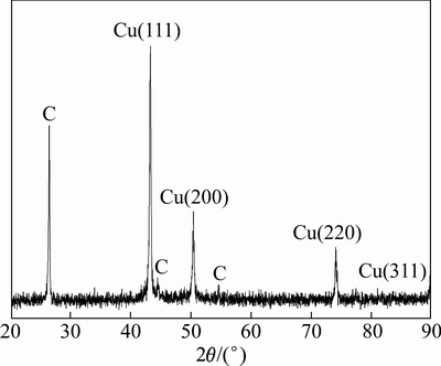
ͼ1 ̼����ͭ��������XRD��
Fig. 1 XRD pattern of Carbon-encapsulated Cu Nanoparticles
2.2 ��Ʒ����ò
ͼ2��ʾΪ̼����ͭ���������͵�TEM��ͼ2(a)�п��Կ������ֿ�������ò���ֲ���������λ������Σ���Ե�Ƚ���������ʾ����ȵijɺ˻��ơ�ͭ������������ȫ���Dz�̼�������������Ե���������ĺ˿ǽṹ���������ɢ�����ã��������������������֮������̼������Ĵ��ڣ����������ӵı����ܣ��Ӷ�����������֮����žۡ���������Ƚϴֲڣ���Ե������������һ�����Ŀ�϶�������ߴ���Ҫ�ֲ���20~100 nm��Χ�ڣ�ƽ������ԼΪ50 nm��ͬʱע���������֮����ճ����������ž���һ������ɳ���״������������״�������������������ܵ������Ӽ�ľ������ͱ���������ͬ���ý����
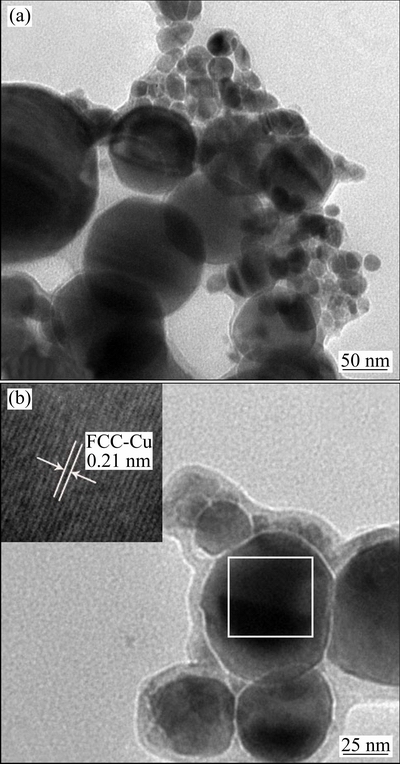
ͼ2 ̼����ͭ��������TEM��HRTEM��
Fig. 2 TEM (a) and HRTEM (b) images of carbon- encapsulated Cu nanoparticles
��ͼ2(b)�п��Կ�����̼����ͭ���������е��͵����-�ں˰����ṹ�������ڲ�������ɫ�����Ϊͭ�����������dzɫ�ı�����̼����Ĥ��̼�������ͭ�ں�֮��Ľ��������������������DZ�̼Ĥ���ܰ������ڲ������˵�ƽ��ֱ��Ϊ40 nm���ң���XRD����ý�������Ǻϣ����̼��������ԼΪ10 nm�����������������γɵĽ����ڽ�����Χ��̼����������У��Dz�̼�ľ����̶Ƚϸߡ���һ������Ʒ�����ں˾ֲ��߱��Ŵ�ɹ۲쵽�����ľ������ƣ���һ���������ڳʹ������У�������ȱ�ݣ�˵���������ᾧ���̶����á����������侧�����ԼΪ0.21 nm����FCC�ṹͭ��(111)������(PDF No.04-0836)�ӽ���Ҳ��XRD�Ľ�������
2.3 ��Ʒ�ĵ�������-�Ѹ�������
�������ڲ�����������������������������ƽ�⣬���ھ���������ͬ����ʣ��ı�������������������������������Ӵ�ʱ����Ϊ���������������Һ��������(78 K)��N2�ڿ��������ϵ�����������������
ͼ3��ʾΪ̼����ͭ��������N2����-�Ѹ������ߣ��˵��������ڵ��͵�Langmuir IV�͡������ѹ��p/p0��0.6�����䷶Χ�ڣ�̼����ͭ��������N2�����������ѹ�������������dz�ƽ���������߱Ƚ�ƽ̹������Kelvin��ʽ���װ뾶С��1.5 nmʱ��������ëϸ������������ˣ��ڵ�ѹ�����������٣�û�г��ֹյ㣬�������Ѹ����ߺܽӽ��������غϡ������̼����ͭ����������(��2 nm)�����ϸߣ��ص����ߵ���ʼ����������Ҫ���������У�ֻ�����ڱ����γɱ��㣬�������о����Ʊ�����Ʒ���п��ֲ���һ�Ľṹ�ص㡣�����ѹ��p/p0��0.6ʱ���������������ѹ��������ʶ����������������������Ѹ��ߺ������ߴ����ͺ����������Ե�IV���ͺ�������Ʒ��������һ�����Ľ��(2~50 nm)�ṹ�ͽϴ�Ŀ�϶�ṹ������������������ëϸ��������IJ�����ʹN2�����ڵ��ڳ�ѹ����������˽�������ڿ�ʼ����ëϸ����ʱ���ڿױ��ϵĻ�״����ĤҺ���Ͻ��У����Ѹ��Ǵӿڵ���������Һ�濪ʼ���Ӷ����Ѹ������߲����غϣ��γ�һ���ͺ�
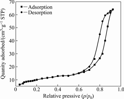
ͼ3 ̼����ͭ�������������Ѹ�����
Fig. 3 Adsorption-desorption curve of carbon-encapsulated Cu nanoparticles
2.4 ��Ʒ�ıȱ����
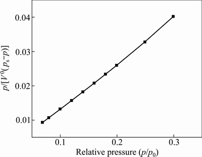
����������������Ʒ�ȱ������ͨ�������Զ�����ʽ��з�ѡ�����������ⶨ��N2�ڹ�������������������N2�����ѹ��p/ps��p/psһ��ѡ����0.05~0.35�ڣ�����BET��ʽ
 (1)
(1)
ʽ�У�pΪʵ��N2ѹ��ֵ��psΪҺ���¶���N2�ı�������ѹ��V0Ϊһ�����ѹ���µ��������������CΪ������VmΪ�����Ӳ㱥��������������ݶ������������������p/psΪ�������ᣬp/[V0(ps-p)]Ϊ�������ᣬ�õ���Ӧ��BET��������ֱ�ߣ���ͼ4��ʾ������ͼ�ⷨ�����Իع鷨���ֱ�ߵĽؾ�b��б��k�ֱ�Ϊb=1/(VmC)��k=(C-1)/(VmC)�����ɴ˵���������������Vm��BET����C�ֱ�ΪVm=1/(k+b)��C=k/b+1�����õ���������ÿ��������һ�������ĵ�������ռ�е�ƽ������õ���λ�������۵ıȱ����(m2/g)��
 (2)
(2)
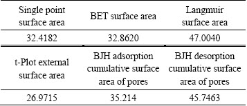
ʽ�У���ΪN2���ӽ������16.2��10-20 m2��NΪ�����ӵ�������6.23��1023������/mol��V0Ϊ�����Ħ�������22.4��103 mL/mol��MΪ����������������g�����Խ�����1��ʾ����ͬ�IJ��Է��������ݵĻ���ʵ��ԭ����ͬ����˵õ��ıȱ������һ���IJ������BET��������������������ĩ��λ�����ıȱ����SMΪ33 m2/g������������Ϊʵ���ұ���⻬�����ο������ù�ʽd=6/(�ѡ�SM)�����У���Ϊ�����ܶȣ�dΪ�����������������Ʒ������ĵ�������dΪ45 nm����õ�BET������TEM��XRD����������һ���IJ�ֵ���������������Ʒ���ֿ��������ճ�����ž����������µġ�
ͼ4 ̼����ͭ��������BETͼ
Fig. 4 BET plots of Carbon-encapsulated Cu nanoparticles
��1 ̼����ͭ�������ıȱ����
Table 1 Specific surface area of carbon-encapsulated Cu nanoparticles (m2/g)
2.5 ��Ʒ�Ŀ��ֲ�
����BJH�����õ�̼����ͭ��������N2�Ѹ����ֲ���ͼ5��ʾ����ͼ5��֪����Ʒ�Ŀ���Ҫ�ֲ���5~20 nm���䷶Χ�ڣ���15 nm������һ�����Է�ֵ��˵��ֱ��Ϊ15 nm�Ŀ�����Ʒ��ռ�ı��������Ʒ���ֲ����߽�խ��˵����Ʒ���ֲ����Ƚϼ��У���ҪΪ�ͽ�ף���ƽ����Ϊ15 nm����Ʒ�����IJ���ԭ��һ�������Դ��̼�ǰ��������ף����ڱ���ᾧȱ����ɾֲ��ṹ��Ԫ�ƻ������ֿṹ����һ����Ҳ�����Dz�ͬ�������ѻ����ɵľۼ����ڵĿ�϶����ɵġ�����BJH����ģ�͵õ�̼����ͭ��������BJH�����ۻ��ܿ��ݻ����Ѹ��ۻ��ܿ��ݻ���Ϊ0.112 cm3/g��BJH����ƽ�������Ѹ�ƽ�����ֱ�Ϊ13��10 nm��
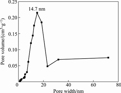
ͼ5 ̼����ͭ��������BJH�Ѹ����ֲ�
Fig. 5 BJH desorption pore size distribution of carbon- encapsulated Cu nanoparticles
3 ����
1) ֱ���绡�������巨�Ʊ���̼����ͭ�������������Եĺ˿ǽṹ���ں�ΪFCC�ṹ��ͭ����������Dz�Ϊ̼����Ĥ��
2) ̼����ͭ����������ò�����ӳ����λ������ηֲ��������ֲ���20~100 nm��Χ��ƽ������Ϊ50 nm���ں˽�����ֱ��Ϊ40 nm���Dz�̼Ĥ���Ϊ10 nm��û�����Ե��ž�����
3) ����ëϸ���������ã���Ʒ��N2����-�Ѹ������߳����ͺ������ͣ�BET������ʽ�õ���Ʒ�ıȱ����Ϊ33 m2/g����������Ϊ45nm����TEM��XRD��õĽ������һ�¡�BJH����ģ�ͼ�����Ʒ�������ۻ��ܿ�����BJH����ƽ�����ֱ�Ϊ0.112 cm3/g��13 nm��
REFERENCES
[1] LOUIE S G. Nanoparticles behaving oddly[J]. Nature, 1996: 384(6610): 612-613.
[2] GRANATA G, YAMAOKA T, PAGNANELLI F, FUWA A. Study of the synthesis of copper nanoparticles: the role of capping and kinetic towards control of particle size and stability[J]. Journal of Nanoparticle Research, 2016, 18(5): 1-12.
[3] Ҷ���, �̼̹�, ���ų�, ���. ������ע���Ʊ�������ͭ���ڵ��罺�е�Ӧ��[J]. �й���ɫ����ѧ��, 2015(5): 1264-1269.
YE Nan-min, CHENG Ji-gui, CHEN Wen-chao, LI Jian-feng. Application of nano-sized copper powders prepared by gel-casting method in conductive adhesives[J]. The Chinese Journal of Nonferrous Metals, 2015(5): 1264-1269.
[4] RESKE R, MISTRY H, BEHAFARID F, ROLDAN CUENYA B, STRASSER P. Particle size effects in the catalytic electroreduction of CO2 on Cu nanoparticles[J]. Journal of the American Chemical Society, 2014, 136(19): 6978-6986.
[5] RANFAGNI A, MUGNAI D. Macroscopic quantum tunneling in differently loaded Josephson junctions[J]. Physics Letters A, 2014, 378(32/33): 2456-2460.
[6] WU D, HUANG L, QUAN X, PUMA G L. Electricity generation and bivalent copper reduction as a function of operation time and cathode electrode material in microbial fuel cells[J]. Journal of Power Sources, 2016, 307: 705-714.
[7] MIN H, LEE B, JEONG S, LEE M. Laser-direct process of Cu nano-ink to coat highly conductive and adhesive metallization patterns on plastic substrate[J]. Optics and Lasers in Engineering, 2016, 80: 12-16.
[8] CHENG Yin, WANG Shou-ling, WANG Ran-ran, SUN Jing, GAO Lian. Copper nanowire based transparent conductive films with high stability and superior stretchability[J]. Journal of Materials Chemistry C, 2014, 2(27): 5309-5316.
[9] LIU Y, WIEK A, DZHAGAN V, HOLZE R. Improved electrochemical behavior of amorphous carbon-coated copper/CNT composites as negative electrode material and their energy storage mechanism[J]. Journal of the Electrochemical Society, 2016, 163(7): A1247-A1253.
[10] SAITO Y. Nanoparticles and filled nanocapsules[J]. Carbon, 1995, 33(7): 979-988.
[11] �� ��, ����Զ, ������, Ҷ��ï, ������, ������. ������ͭ����ըҩ����ϳ�ʯī����ͭ������[J]. ϡ�н��������빤��, 2014, 43(2): 465-469.
LUO Ning, LIU Zhi-yuan, CHEN Tian-wu, YE Lin-mao, LIU Kai-xin, LI Xiao-jie. Metal copper ions doped explosives for synthesis of graphite coated copper nanoparticles[J]. Rare Metal Materials and Engineering, 2014, 43(2): 465-469.
[12] ����Ƽ, �ź���, �� ��, �ӽ�ɽ, �ش���, �� п. ̼��������ͭ���ӵ��Ʊ�������������[J]. �й���ɫ����ѧ��, 2010, 20(9): 1766-1774.
LI Li-ping, ZHANG Hai-yan, LIN Jin, PANG Jin-shan, HE Chun-hua, NING Xin. Preparation and anti-oxidation properties of carbon-coated copper nanoparticles[J]. The Chinese Journal of Nonferrous Metals, 2010, 20(9): 1766-1774.
[13] ZHANG Fan, CUI Lan, LIN Kui, JIN Feng-min, WANG Bin, SHI Shu-xiu, YANG De-an, WANG Hui, HE Fei, CHEN Xiao-ping, CUI Shen. Preparation of carbon-encapsulated iron nanoparticles in high yield by DC arc discharge and their characterization[J]. Journal of Alloys and Compounds, 2013, 553: 367-374.
[14] LUO Ning, LI Xiao-jie, LIU Kai-xin, YE Lin-mao, CHEN Tian-wu. Preparation of carbon-coated copper nanoparticles by detonation decomposition of copper ion doped sol�Cgel explosive precursors[J]. Journal of Nanoparticle Research, 2013, 15(5): 1-9.
[15] KIM C, LEE G, RHEE C, LEE M. Expeditious low-temperature sintering of copper nanoparticles with thin defective carbon shells[J]. Nanoscale, 2015, 7(15): 6627-6635.
[16] LI Yong-sheng, SHI Jian-lin. Hollow-structured mesoporous materials: Chemical synthesis, functionalization and applications[J]. Advanced Materials, 2014, 26(20): 3176-3205.
[17] YANG Sheng-chun, LUO Xiao. Mesoporous nano/micro noble metal particles: Synthesis and applications[J]. Nanoscale, 2014, 6(9): 4438-4457.
[18] GU D, JIA C, WEIDENTHALER C, BONGARD H, SPLIETHOFF B, SCHMIDT W, SCHU?TH F. Highly ordered mesoporous cobalt-containing oxides: structure, catalytic properties, and active sites in oxidation of carbon monoxide[J]. Journal of the American Chemical Society, 2015, 137(35): 11407-11418.
[19] THOMMES M, CYCHOSZ K A. Physical adsorption characterization of nanoporous materials: progress and challenges[J]. Adsorption-Journal of the International Adsorption Society, 2014, 20(2/3): 233-250.
[20] LI Hui, XIAO De-li, HE Hua, LIN Rui, ZUO Peng-li. Adsorption behavior and adsorption mechanism of Cu(II) ions on amino-functionalized magnetic nanoparticles[J]. The Chinese Journal of Nonferrous Metals, 2013(9): 2657-2665.
[21] KIM K C, YOON T, BAE Y. Applicability of using CO2 adsorption isotherms to determine BET surface areas of microporous materials[J]. Microporous and Mesoporous Materials, 2016, 224: 294-301.
[22] WEI Zhi-qiang, XIA Tian-dong, BAI Li-feng, WANG Jun, WU Zhi-guo, YAN Peng-xun. Efficient preparation for Ni nanopowders by anodic arc plasma[J]. Materials Letters, 2006, 60(6): 766-770.
Specific surface area and pore structure of carbon coated copper nanoparticles
BAI Jun-shan1, WEI Zhi-qiang1, 2, ZHU Xue-liang1, FENG Wang-jun1, YANG Hua1, 2, JIANG Jin-long1
(1. School of Science, Lanzhou University of Technology, Lanzhou 730050, China;
2. State Key Laboratory of Advanced Processing and Recycling of Non-ferrous Metals, Lanzhou University of Technology, Lanzhou 730050, China)
Abstract: Carbon encapsulated Cu nanoparticles were successfully prepared by DC arc discharging plasma technology. The product was characterized by X-ray diffractometry (XRD), high resolution transmission electron microscopy (HRTEM) and low-temperature N2 adsorption desorption to determine the morphology, crystal structure, particle size, specific area and pore structure. The results indicate that the carbon encapsulated copper nanoparticles have typical core shell structure, the core is metal face centered cubic structure copper, and the outer shell is graphite carbon layer. The particles morphology mainly exhibit spherical or ellipsoidal shapes with relatively uniform particle size and good dispersion. The particle size distribution is in the range of 20-100 nm, the average particle size is 50 nm, and the thickness of the shell carbon layer is about 10 nm. The N2 adsorption and desorption isotherm belongs to type ��, most of pores between grains are mesoporous pore, the BET specific surface area is 33 m2/g, and the equivalent particle size is 45 nm, which is consistent with the results measured by TEM and XRD. The BJH adsorption cumulative pore volume and adsorption average pore size are 0.112 cm3/g and 13 nm, respectively.
Key words: carbon-encapsulated; core-shell structure; nitrogen adsorption; specific surface area; pore structure
Foundation item: Project(51261015) supported by the National Natural Science Foundation of China; Project(1308RJZA238) supported by the Natural Science Foundation of Gansu Province, China
Received date: 2016-06-17; Accepted date: 2016-11-24
Corresponding author: WEI Zhi-qiang; Tel: +86-931-2973780; E-mail: zqwei7411@163.com
(�༭ ��ѧ��)
������Ŀ��������Ȼ��ѧ����������Ŀ(51261015)������ʡ��Ȼ��ѧ����������Ŀ(1308RJZA238)
�ո����ڣ�2016-06-17�������ڣ�2016-11-24
ͨ�����ߣ�κ��ǿ�����ڣ���ʿ���绰��0931-2973780��E-mail��zqwei7411@163.com
ժ Ҫ������ֱ���绡�ŵ�������弼���Ʊ�̼����ͭ��������������Ʒ����ò������ṹ�����ȡ��ȱ�����Ϳṹ���ø߷ֱ����������(HRTEM)��X����������(XRD)��N2��-�Ѹ��Ȳ����ֶν��з��������������ֱ���绡�������弼���Ʊ���̼����ͭ���������е��͵ĺ˿��ͽṹ���ں�Ϊ���������Ľ���ͭ�����Ϊʯī̼�㡣������Ҫ�����λ������Σ�������ԱȽϾ��ȣ���ɢ�����ã������ֲ���20~100 nm��Χ�ڣ�ƽ������Ϊ50 nm�����̼��ĺ��Ϊ10 nm��̼����ͭ�������ĵ����������������ͣ�����֮��Ŀ�϶�Խ��Ϊ������Ʒ��BET�ȱ����Ϊ33 m2/g����������Ϊ45 nm����TEM��XRD��õĽ������һ�¡�BJH�����ۻ��ܿ�����BJH����ƽ�����ֱ�Ϊ0.112 cm3/g��13 nm��
