
DOI: 10.11817/j.issn.1672-7207.2018.08.006
���-2-��ȩ��ϣ���ͭ�����ĺϳɼ����������
���磬�վ�������ޱ�����ٴ�
(���ϴ�ѧ ��ѧ�뻯��ѧԺ������ ��ɳ��410083)
ժ Ҫ��
��-2-��ȩ���鰷ϣ���ͭ(II)�����[Cu(Quin-Hist)Cl2]������Ԫ�ط����ͺ����������б�����ͨ��X�ߵ�������ȷ���侧��ṹ���о�����������þ������ڵ�б���壬P21/n�ռ�Ⱥ����������Ϊa= 0.858 90(7) nm��b=1.282 38(10) nm��c=1.459 76(12) nm����=99.346 0(10)�㣻�����Ϊ���������˫�ռ乹�ͣ���ͭ(II)���������Сţ����DNA(CT-DNA)֮��ͨ�����۽�ϵķ�ʽ����ã����⣬ͭ(II)������MCF-7�����ٰ�ϸ��ϵ��A-549�˷�Сϸ���ΰ�ϸ��ϵ��SKOV-3���ѳ���ϸ��ϵ���н�ǿ������ϸ�������ԣ�����˳����ȣ�ͭ(II)������MCF-7ϸ��ϵ���и�ǿ������ϸ�������ԡ�
�ؼ��ʣ�
ͭ�������ϣ���������ṹ��DNA�����ϸ����������
��ͼ����ţ�O614.121 ���ױ�־�룺A ���±�ţ�1672-7207(2018)08-1878-06
Synthesis and bio-activity of copper(ii) complex of schiff-base derived from quinoline-2-carboxaldehyde
HU Xi, YAN Jun, FANG Ziwei, ZHANG Shouchun
(School of Chemistry and Chemical Engineering, Central South University, Changsha 410083, China)
Abstract: A novel copper(II) complex, i.e. [Cu(Quin-Hist)Cl2] (Quin-Hist=2-(4-imidazolyl) ethylimino-quinoline), was prepared and structurally characterized by elemental analysis and IR spectra. The results show that the complex crystallized belongs to monoclinic space group P21/n with a=0. 858 90(7) nm, b=1.282 38(10) nm, c=1.459 76(12) nm, ��=99.346 00(10)��. The copper(II) ion is situated in a trigonal bipyramid geometry. There is apparent interaction between the copper(II) complex and calf thymus DNA(CT-DNA) via a groove binding mode. Moreover, the copper(II) complex exhibits in vitro cytotoxicity against MCF-7,A549 and SKOV-3 cell lines. Especially, the copper(II) complex shows higher growth inhibitory rates than cisplatin against MCF-7 cell line.
Key words: copper complex; schiff-base; crystal structure; DNA binding; cytotoxicity
��֢�ǵ��µ��������������ߵļ���֮һ����������в�����ǵ����彡����˳����Ŀǰ�ٴ��ϳ��õ�һ�ֿ���ҩ��������صĶ������á���ҩ�Լ��Ѵ�л��ȱ�������������ٴ�ʹ�ã���ˣ��з�����Ч���Ͷ���ˮ���Ժá�������Χ�������С���ӽ���������ܵ�������Խ��Խ��Ĺ�ע[1-4]��ͭ������������Ԫ��֮һ���������ڵ����ʺ�ø����Ҫ��ɲ��֣���������ϵ�о�����Ҫ���á��벬��ȣ�ͭ���и��ߵ���������Ժ��͵Ķ��ԣ���ˣ�ͭ����ﱻ��Ϊ�Ǽ̲�������������Ӧ��ǰ���Ŀ���ҩ��֮һ[5]�������������Ǻϳ��˴���ͭ����ﲢ������Щ�������н�ǿ�Ŀ�������[6]������Щ������У�ϣ�����ͭ�������о���Ϊ����עĿ������ϣ�����ﱾ������һ����������ԣ�ϣ��������Ҳ��֤�����н�ǿ�Ŀ����������뿹������[7-8]��ZHANG��[9-10]�ѱ���������-2-��ȩ����������ϣ���������Щ��������DNA��ϣ��������и�DNA���Ҷ�����ϸ���н�ǿ�������������á�Ϊ�˽�һ���о����-2-��ȩ��ϣ������������������ԣ���������ѡ���Ծ���������Ե��鰷�����-2-��ȩ��Ϊԭ�Ϻϳ�ϣ������壬�����ϳ�ͭ(II)����������ǿͭ��������������ԣ��Ӷ���ǿ������������ԡ�
1 ʵ��
1.1 ʵ���Լ�������
��Ҫ�Լ�Ϊ�����-2-��ȩ���������飬���Ǽ���������(Tris)���廯�Ҷ�(EB)��������Sigma��˾��Сţ����DNA(CT-DNA)������MBI Fermentas��˾��ˮ��Ϊ��������ˮ�������Լ�����Ϊ��������
��Ҫ������Bruker VEC-TOR22 ���������(KBrѹƬ)��Perkin-ElmerԪ�ط����ǣ�Shimadzu UV-2450 ����-�ɼ��ֹ��ȼƣ�Hitachi F4600ӫ������ǣ�Jasco J-815Բ��ɫ�����ǣ�Bruker CCD X�������ǡ�
1.2 �����[Cu(Quin-Hist)Cl2]�ĺϳ�
��0.157 g(1.0 mmol)���-2-��ȩ��0.184 g(1.0 mmol)�������鰷����15 mL�Ҵ��У��μӼ���NaOH��Һ���������2 h��Ȼ��μӺ�0.085 g(0.5 mmol) CuCl2��2H2O���Ҵ���Һ5 mL����������4 h����ȴ�����º���ˣ�����ɫ�������Ҵ�������ϴ�����Ρ����ڿ����и��Ȼ��������ɫ����������ˮ�Ҵ��У����ûӷ���������ɫ��״��������������Ϊ0.137 g(����Ϊ71.4%)����C15H14N4CuCl2Ԫ�������������з���������ֵ���£�C��������Ϊ46.91%��HΪ3.73%��NΪ14.45%��ʵ��ֵ(��������)���£�CΪ46.84%��HΪ3.70%��NΪ14.51%������ͼ����(KBrѹƬ��cm-1)������£�3 111 cm-1���ķ�Ϊ�����ϵ�N��H���������շ壻1 654 cm-1���ķ�Ϊ������ϵ�C=N���������շ壻1 592 cm-1���ķ�ΪC=N���������շ塣
1.3 �����[Cu(Quin-Hist)Cl2]����ṹ�ⶨ
����Bruker CCD�������ռ�ͭ����ᄃ���X���������ݡ����¶�293 K�£������ԴΪʯī��ɫ����ɫ����Mo K�� ���ߣ���5.0~50.0 min-1 ���ٶ����æ�-2�� ɨ�跽ʽ�ռ���������������������������-�������ӺͰ뾭������У����������ԭ�����������ۼ���ȷ������ȫ������ԭ�����꼰����������Ȳ�������ȫ������С���˷����������м�����SHELXTL������ɡ������ľ���ѧ��������1��
1.4 �����[Cu(Quin-Hist)Cl2]��DNA�������
��Tris-HCl������Һ(5 mmol/L Tris��5 mmol/L HCl��50 mmol/L NaCl��pH=7.4)�ܽ�һ������CT-DNA���ⶨ��DNA��Һ��260 nm��280 nm��������ȣ���2������ȱ�ֵԼΪ1.9��������DNA��Һ�в����е�����[11]��DNA��Һ��Ũ�ȿ�ͨ��260 nm������Ȧ�260ȷ��(��260=6 600 L��mol-1��cm-1)[12]��
ȡһ����[Cu(Quin-Hist)Cl2]��������DMSO�У�Ȼ���������Tris-HCl������Һϡ����Ũ��Ϊ5.0��10-5 mol/L��ȡ�������Һ��DNA��Һ����ͬ���ʵ���֮��(r[n(Cu)/n(DNA)] Ϊ 0��0.2��0.4�� 0.6)��ϣ���������-�ɼ��ֹ��ȼƷֱ�ⶨ�����������Һ������-�ɼ����ס�ͭ�������DNA�Ľ�ϳ��� kb ������ʽ����ó�[13]��
c[DNA]/(��a �C ��f)=c[DNA]/(��b�C��f)+1/[kb(��b �C ��f)] (1)
ʽ�У�c[DNA]Ϊ��DNA��Ũ�ȣ���aΪ�����ı���Ħ������ϵ������fΪ��������Ħ������ϵ������bΪ�������DNA��ֽ�Ϻ��Ħ������ϵ����kbΪ��ϳ���������c[DNA]/(��b-��f)��c[DNA]��ͼ�õ���ֱ��б����ؾ�ı�ֵ��
��1 �����[Cu(Quin-Hist)Cl2]�ľ���ѧ��������ֵ
Table 1 Crystal and structure refinement parameters and their values for complex [Cu(Quin-Hist)Cl2]

��CT-DNA(1.0��10-4 mol/L)��EB(1.0��10-5 mol/L)�����Һ�л����μ�[Cu(Quin-Hist)Cl2]��Һ(5.0��10-5 mol/L)���������²ⶨÿ�μ�����ӫ����ı仯(��������Ϊ530 nm�����䲨��Ϊ600 nm)�������k����Stern-Volmerʽ����ó�[14]��
I0/I=1+k��r[n(Cu)/n(DNA)] (2)
ʽ�У�I0��I�ֱ�Ϊ�����������ʱӫ����ķ���ǿ�ȣ�r[n(Cu)/n(DNA)]Ϊͭ�������DNA�����ʵ���֮�ȡ���I0/I��r[n(Cu)/n(DNA)]��ͼ�������kΪ��ֱ�ߵ�б�ʡ�
��25 ��ⶨ[Cu(Quin-Hist)Cl2]��DNA(1.0��10-4 mol/L)�Բ�ͬ���ʵ���֮�Ȼ��������Һ��Բ��ɫ���ס�ɨ�貨��Ϊ220~320 nm��ɨ���ٶ�Ϊ10 nm/min��
1.5 ����ϸ�������Բ���
�����[Cu(Quin-Hist)Cl2]������ϸ��������ѡ�������ٰ�ϸ��ϵ(MCF-7)���˷�Сϸ���ΰ�ϸ��ϵ(A-549)�����ѳ���ϸ��ϵ(SKOV-3)����MTT(3-(4,5-dimethylthiazol-2-yl)-2,5-diphenyltetrazolium bromide)�����в��ԣ�����˳��Ϊ���Զ��ա�����ϸ����96�װ������������Ϊ5%��CO2��37 ������20 h��ϸ�����ں��뺬�в�ͬŨ���ݶȵ�ͭ����������Һ100 ��L����48 h��Ȼ����ÿ���м��뺬MTT��PBS������Һ(20 ��L��5 g/L)������4 h�������ÿ���м���200 ��L DMSO��ҡ��10 min������ø���Ǽ�⡣
2 ���������
2.1 ����ṹ
���-2-��ȩ��������鰷�����ʵ�����Ϊ1:1�ı�����Ϸ�Ӧ����ϣ�����Ȼ�����CuCl2��2H2O��ͨ��ԭλ��Ӧ�õ���������[Cu(Quin-Hist)Cl2]��[Cu(Quin-Hist)Cl2]�ķ��ӽṹ��ͼ1��ʾ����Ҫ�����ͼ������2��ʾ�������������ļ����ͼ������3��ʾ������������Ľ���Cu(II)���Ӵ��ڱ�������˫��λ�����У����Ϊ0.65(��=(��-��)/60�����У���ΪCl(2)-Cu(1)-Cl(1)�ļ��ǣ���ΪN(1)-Cu(1)-N(3)�ļ��ǣ����ͦ·ֱ�Ϊ130.8���169.8��)������Ϊ0��1ʱ�������ֱ�Ϊ������ķ�������˫���ͣ�����Ϊ0~1ʱ�������Ϊ��������˫����)[15]���������N(1)ԭ�Ӻ�������N(3)ԭ��λ������˫��2�����ᶥ�㣬�ǰ���N(2)ԭ�Ӻ�2����ԭ���������ƽ�档����ƽ����N(2)-Cu(1)-Cl(1)��N(2)-Cu(1)-Cl(2)��Cl(2)-Cu(1)-Cl(1)�ļ��Ǿ�����ȣ���ֵ�ֱ�Ϊ106.2(6)�㣬122.5(6)���130.8(3)�㡣Cu(1)-Cl(1) ��Cu(1)-Cl(2)�ļ���Ҳ����ȣ���ֵ�ֱ�Ϊ0.241 0(6) nm��0.229 4(7) nm��������N(1)-Cu(1)-N(3)�ļ���Ϊ169.8(7)�㣬�ȹ��������˫�еĸü���180���С��������Cu(1)-N(1)��Cu(1)-N(3)�ļ����ֱ�Ϊ0.203 9(17) nm��0.196 5(17) nm��������ƽ����Cu(1)-N(2)����(0.207 4(18) nm)���̡����������һЩ������Cu(II)������е�Cu-N���������������[16]��������һЩϣ���ͭ(II)�������Cu-N�ǰ��������Գ�[17-18]��[Cu(Quin-Hist)Cl2]�ľ���ṹ�ѻ�ͼ��ͼ2��ʾ�����Ӽ���������N(4)������Cl(1)��D������A����Ϊ0.315 0 nm������֮�⣬������е�����������ڵ�����֮����ڦ�-�� �ѻ����ã�����ƽ��ļ��Ϊ0.331 5(3) nm�������ͨ������ͦ�-�жѻ������γɾ�����״�ṹ�ij�������ϵ��
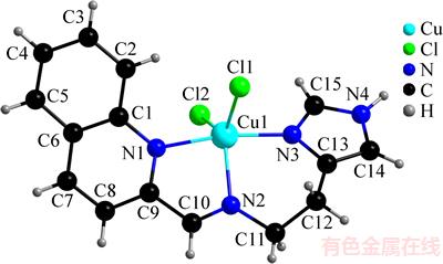
ͼ1 �����[Cu(Quin-Hist)Cl2]�ķ��ӽṹ
Fig. 1 Molecular structure of [Cu(Quin-Hist)Cl2]
��2 ��������Ҫ��������Ҫ����
Table 2 Selected bond distances (nm) and angles (��) of [Cu(Quin-Hist)Cl2]

��3 ������е�����ļ����ͼ���
Table 3 Hydrogen bond lengths (nm) and bond angles (��) of [Cu(Quin-Hist)Cl2]


ͼ2 ��������ͨ������ͦ�-�жѻ��γɵ�һά��������״�ṹ
Fig. 2 [Cu(Quin-Hist)Cl2] crystal structure with relevant one-dimensional supramolecular structure formed by hydrogen-bonding and ��-�� stacking interactions
2.2 �������DNA����÷�ʽ���о�
2.2.1 �������չ���
DNA���ձ���Ϊ�������ҩ�����Ҫ���ðе㣬�о������������DNA֮�������ö��ڿ�������Ч�Ŀ����Լ�������Ҫ���塣������������-�ɼ����չ��ס�ӫ�������Բ��ɫ���ȷ����������[Cu(Quin-Hist)Cl2]��DNA֮������ý����о�����С���ӽ����������Ƕ�뷽ʽ��DNA����Խ��ʱ�����������չ�����ַ�λ���ƺͼ�ɫЧӦ���������չ��ױ仯�̶������������йء���ɫЧӦԽ��˵�������������DNA����Խǿ����Ƕ�뷽ʽ��ϵĿ�����Խ�ߡ����շ����Խ����˵���������DNAǶ��̶�Խ��[19]���������DNA���õ��������չ�����ͼ3��ʾ����ͼ3�ɼ�������DNAŨ�����ӣ��������������չ�����251 nm�������˼�ɫЧӦ����ɫ��ԼΪ8.42%����û�г��ֺ�������Ϊ�˶����Ƚ�ͭ�������DNA�������������ʽ(1)����������DNA�Ľ�ϳ���kbΪ6.70��104 mol/L���ó���ԶС�ھ����Ƕ���Լ�EB�Ľ�ϳ���(EB-DNA��ϳ���Ϊ1.00��106 mol/L)[20]��˵����ͭ�������DNA�Ľ��������EB��DNA�Ľ�������ͣ��Ҹ�����������ͨ���Ǿ�������÷�ʽ�繵�۷�ʽ��DNA���[21]��

ͼ3 ��ͬŨ���������DNA����õ��������չ���
Fig. 3 Electronic absorption spectra of complex (5.0��10-5 mol/L) absence (a) and presence (b-d) of increasing amounts of CT-DNA at the ratio r=0, 0.2, 0.4, 0.6
2.2.2 ӫ�����
DNA��EB������ӫ���Զ�����������EB���뵽DNAС�����ļ���Ժ���ӫ��ǿ�Ⱦͻ�����ǿ����EB��DNA��������������ʱ��ӫ��ǿ���ֻ����������ˣ����Ը���������EB-DNA������ӫ��ǿ�ȵ�Ӱ�����о����������DNA�������á������[Cu(Quin-Hist)Cl2]��DNA����õ�ӫ�������ͼ4��ʾ(ÿ����EB-DNA��ϵ������20 ��L�������Һ)����ͼ4�ɼ������������Ũ�����ӣ�EB-DNA��������600 nm����ӫ��ǿ��������������Ϊ49%�������ͭ��������ȡ��EB-DNA�������е�EB����DNA������Ч��ϡ�ͭ�����������Թ��۷�ʽ��DNA���[22]�������������չ��ײⶨ���һ�¡�
ͼ4 ��ͬ����������EB-DNA��ϵ����õ�ӫ�����ͼ
Fig. 4 Fluorescence emission spectra of ethidium bromide (EB)-CT-DNA system in absence and presence of complex
2.2.3 Բ��ɫ��
DNA�Ǿ���˫�����ṹ�����Է��ӣ���Բ��ɫ���ϻ����2���壺275 nm���������245 nm���ĸ��塣��������DNA����Ԧ�-�жѻ��γɵģ�����������ζ�ż���ѻ���ʵ����������DNA�������Բ����ģ�����������ζ��������DNA�н�������[23]�������[Cu(Quin-Hist)Cl2]��DNA����õ�Բ��ɫ����ͼ5��ʾ����ͼ5�ɼ�������r[n(Cu)/n(DNA)]����Բ��ɫ����275 nm��������ǿ��������245 nm���ĸ���ǿ��������С������û�з����״����ƻ����ơ������ͭ�������DNA��������ã�����DNA�����������Ա仯����247 nm�������½�������������������DNA�����������ã�ʹDNAת��ΪA����[24]��

ͼ5 ��ͬŨ���������DNA����õ�Բ��ɫ��ͼ
Fig. 5 CD spectra of CT-DNA in absence and presence of increasing amounts of complex at the ratio r
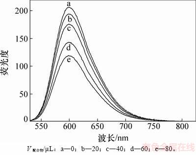
2.3 ����ϸ�������о�
IC50Ϊɱ����������ϸ�������ҩ��Ũ�ȣ���ֵԽ�ͣ���ҩ���ϸ������Խ�ߡ�������˳����Ϊ���Զ��գ�ͨ��MTT�����������[Cu(Quin-Hist)Cl2]��3�ֲ�ͬ����ϸ��ϵ(MCF-7��A-549��SKOV-3)��ϸ�������ԣ������ͼ6��ʾ����ͼ6�ɼ�����ͭ������MCF-7��A-549��SKOV-3��3��ϸ��ϵ��IC50�ֱ�Ϊ15.2��27.2��25.8 ��mol/L���ɴ�˵����������MCF-7ϸ��ϵ������ǿ������ϸ�������ԣ�������2��ϸ��ϵҲ�в�ͬ�̶ȵ������������á����⣬��ͭ������MCF-7ϸ��ϵ��IC50��˳���ĵ�(IC50=20.4 ��mol/L)�������ֳ�ǿ��˳��������ϸ�������ԣ�����������[Cu(Quin-Hist)Cl2]������չ��Ϊһ��DZ�ڵĿ����Լ���

ͼ6 ������3�ְ�ϸ�����ϸ��������ͼ (��˳��Ϊ����)
Fig. 6 Cytotoxic activity of [Cu(Quin-Hist)Cl2] against three human tumor cell lines (cisplatin is used as positive control)
3 ����
1) �����-2-��ȩ���������鰷��CuCl2��2H2OΪԭ�ϣ�ͨ��ԭλ��Ӧ�ϳ���һ���µ�ϣ�����ͭ�����[Cu(Quin-Hist)Cl2]��
2) ��ϣ�����ͭ�����ṹΪ���������˫���ͣ�����������ͨ������ͦ�-�жѻ������γɾ�����״�ṹ�ij�������ϵ��
3) �����[Cu(Quin-Hist)Cl2]�Թ��۽�ϵķ�ʽ��CT-DNA��ϣ����Ҷ�MCF-7ϸ��ϵ���б�˳����������ϸ�������ԣ���������������Ϊһ��DZ�ڵĿ����Լ���
�ο����ף�
[1] GARBUTCHEONSINGH K B, GRANT M P, HARPER B W, et al. Transition metal based anticancer drugs[J]. Current Topics in Medicinal Chemistry, 2011, 11(5): 521-542.
[2] OLIVERI V, VECCHIO G. 8-Hydroxyquinolines in medicinal chemistry: a structural perspective[J]. European Journal of Medicinal Chemistry, 2016, 120(14): 252-274.
[3] �����, ��ٻٻ, ����Ʒ, ��. 8-�ǻ��������������������������Ϳ��������о���չ[J]. �й���ѧ: ��ѧ, 2017, 47(2): 172-182.
QIN Jiaolan, CAO Qianqian, QIN Qipin, et al. Progress on antitumor and antibacterial activity of 8-hydroxyquinoline and its derivatives metal complexes[J]. Scientia Sinica Chimica, 2017, 47(2): 172-182.
[4] QIN Qipin, CHEN Zhenfeng, QIN Jiadan, et al. Studies on antitumor mechanism of two planar platinum(II) complexes with 8-hydroxyquinoline: synthesis, characterization, cytotoxicity, cell cycle and apoptosis[J]. European Journal of Medicinal Chemistry, 2015, 92(6): 302-313.
[5] MARZANO C, PELLEI M, TISATO F, et al. Copper complexes as anticancer agents[J]. Anti-Cancer Agents in Medicinal Chemistry, 2009, 9(2): 185-211.
[6] ZHANG Zhen, WANG Huiyun, YAN Maocai, et al. Novel copper complexes as potential proteasome inhibitors for cancer treatment (Review)[J]. Molecular Medicine Reports, 2017, 15(1): 3-11.
[7] LIAN Wenjing, WANG Xintian, XIE Chengzhi, et al. Mixed-ligand copper(ii) Schiff base complexes: the role of the co-ligand in DNA binding, DNA cleavage, protein binding and cytotoxicity[J]. Dalton Transactions, 2016, 45(22): 9073-9087.
[8] SHEBLL M. Synthesis,spectroscopic characterization and antimicrobial activity of binuclear metal complexes of a new asymmetrical Schiff base ligand: DNA binding affinity of copper(II) complexes[J]. Spectrochimica Acta Part A: Molecular and Biomolecular Spectroscopy, 2014, 117(1): 127-137.
[9] ZHANG Shouchun, DONG Juanjuan, FAN Xiaorui, et al. Cobalt(II) complexes with thiosemicarbazone as potential antitumor agents:synthesis,crystal structures, DNA interactions, and cytotoxicity[J]. Spectrochimica Acta: Part A (Molecular and Biomolecular Spectroscopy), 2013, 66(24): 4268-4279.
[10] ZHANG S C, DONG J J, FAN X R, et al. A new nickel(II) complex with the thiosemicarbazone of quinoline-2- carboxaldehyde: structure, DNA-binding, cleavage, and cytotoxic activities[J]. Journal of Coordination Chemistry, 2012, 65(17): 3098-3110.
[11] MARMUR J. A procedure for the isolation of deoxyribonucleic acid from micro-organisms[J]. Journal of Molecular Biology, 1961, 3(1): 208-218.
[12] REICHMANN M E, RICE S A, THOMAS C A, et al. A further examination of the molecular weight and size of desoxypentose nucleic acid[J]. Journal of American Chemical Society, 1954, 76(11): 3047-3053.
[13] WOLFE A, JR SHIMER G H, MEEHAN T. Polycyclic aromatic hydrocarbons physically intercalate into duplex regions of denatured DNA[J]. Biochemistry, 1987, 26(20): 6392-6396.
[14] EFTINK M R, GHIRON C A. Fluorescence quenching studies with proteins[J]. Anal Biochem, 1981, 114(2): 199-227.
[15] ADDISON A W, RAO T N, DEEDIJK J, et al. Synthesis, structure, and spectroscopic properties of copper(II) compounds containing nitrogen-sulphur donor ligands; the crystal and molecular structure of aqua[1,7-bis(N-methylbenzimidazol- 2'-yl)-2,6-dithiaheptane]copper(II) perchlorate[J]. J Chem Soc Dalton Trans, 1984(7): 1349-1356.
[16] SADHUKHAN D, RAY A, DAS S, et al. Effect of ligand substitution on DNA binding ability of two new square planar copper(II)-Schiff base complexes[J]. J Mol Struc, 2010, 975(1): 265-273.
[17] DONG Jianfang, LI Lianzhi, LIU Guihua, et al. Synthesis, crystal structure and DNA-binding properties of a new copper(II) complex with l-valine Schiff base and 1,10-phenanthroline[J]. J Mol Struc, 2011, 986(1/2/3): 57-63.
[18] QIAO Xin, MA Zhongying, XIE Chengzhi, et al. Study on potential antitumor mechanism of a novel Schiff Base copper(II) complex: synthesis, crystal structure, DNA binding, cytotoxicity and apoptosis induction activity[J]. J Inorg Biochem, 2011, 105(5): 728-737.
[19] NAIR R B, TENG E S, KIRKLAND S L, et al. Synthesis and DNA-binding properties of [Ru(NH3)4dppz]2+[J]. Inorganic Chemistry, 1998, 37(1): 139-141.
[20] WARING M J. Complex formation between ethidium bromide and nucleic acids[J]. Journal of Molecular Biology, 1965, 13(1): 269-282.
[21] XU Zhihong, CHEN Fengjuan, XI Pinxian, et al. Synthesis, characterization, and DNA-binding properties of the cobalt(II) and nickel(II) complexes with salicylaldehyde 2-phenylquinoline-4-carboylhydrazone[J]. J Photochem Photobiol A, 2008, 196(2): 77-83.
[22] KRISHNA A G, KUMAR D V, KHAN B, et al. Taxol-DNA interactions:fluorescence and CD studies of DNA groove binding properties of taxol[J]. Biochimica et Biophysica Acta (BBA)-General Subjects, 1998, 1381(1): 104-112.
[23] COLLINS J G, SHIELDS T P, BARTON J K. 1H-NMR of Rh(NH3)4phi3+ Bound to d(TGGCCA)2:classical intercalation by a nonclassical octahedral metallointercalator[J]. Journal of the American Chemical Society, 1994, 116(22): 9840-9846.
[24] CHAUHAN M, BANERJEE K, ARJMAND F. DNA binding studies of novel copper(ii) complexes containing L-tryptophan as chiral auxiliary: in vitro antitumor activity of Cu-Sn2 complex in Human neuroblastoma cells[J]. Inorg Chem, 2007, 46(8): 3072-3082.
(�༭ �²ӻ�)
�ո����ڣ�2017-10-23�������ڣ�2017-12-20
������Ŀ(Foundation item)��������Ȼ��ѧ����������Ŀ(21301194)(Project(21301194) supported by the National Natural Science Foundation of China)
ͨ�����ߣ����ٴ�����ʿ�������ڣ����½�����������������о���E-mail��zhang_shch@sina.cn
ժҪ���ϳ�һ���µ����-2-��ȩ���鰷ϣ���ͭ(II)�����[Cu(Quin-Hist)Cl2]������Ԫ�ط����ͺ����������б�����ͨ��X�ߵ�������ȷ���侧��ṹ���о�����������þ������ڵ�б���壬P21/n�ռ�Ⱥ����������Ϊa= 0.858 90(7) nm��b=1.282 38(10) nm��c=1.459 76(12) nm����=99.346 0(10)�㣻�����Ϊ���������˫�ռ乹�ͣ���ͭ(II)���������Сţ����DNA(CT-DNA)֮��ͨ�����۽�ϵķ�ʽ����ã����⣬ͭ(II)������MCF-7�����ٰ�ϸ��ϵ��A-549�˷�Сϸ���ΰ�ϸ��ϵ��SKOV-3���ѳ���ϸ��ϵ���н�ǿ������ϸ�������ԣ�����˳����ȣ�ͭ(II)������MCF-7ϸ��ϵ���и�ǿ������ϸ�������ԡ�
