
���±�ţ�1004-0609(2016)-11-2319-07
Gd����ע��Թ���̬Mg-Nd-Sr-Zr�Ͻ����︯ʴ��Ϊ��Ӱ��
��ѧΰ1, 2��������1, 2��������1, 2����־��1, 2����ǿʤ1, 2
(1. �Ͼ�����ѧԺ ���Ϲ���ѧԺ���Ͼ� 211167��
2. �Ͼ�����ѧԺ ����ʡ�Ƚ��ṹ������Ӧ�ü����ص�ʵ���ң��Ͼ� 211167)
ժ Ҫ��
�Թ���̬Mg-Nd-Sr-Zr�Ͻ��������(Gd)����ע����Դ���������SRIM 2008������Gd����ע����̽�����ģ����������ù�ѧ����(OM)�۲���þ�Ͻ������֯��������X ����������(XRD)�������Բ��е�������ɣ�ͬʱ���X���߹��������(XPS)�������Բ��л�ѧ�ɷ���Ԫ�ؼ�̬�����õ绯ѧʵ�鼰����ʵ�������˻���þ�Ͻ���ע��þ�Ͻ���ģ����Һ(SBF)�е����︯ʴ��Ϊ�����������Gd����ע����������߹���̬Mg-Nd-Sr-Zr�Ͻ���SBF�������︯ʴ���ܣ��ҵ�ע�����Ϊ2.5��1016 cm-2ʱ��ע��þ�Ͻ�������︯ʴ������ѡ�
�ؼ��ʣ�
��ͼ����ţ�TG174.44��TG146.4���� ���ױ�־�룺A
�˿����仯�����Լ�����Խ������ٵIJ�����������ҽ�ò��ϵ�����Ѹ�����ӡ�þ�Ͻ���Ϊ��һ������ҽ�ò��ϣ����������������ȣ���������ͻ���ŵ�[1-4]��1) þ�Ͻ������������オ���ԣ��ܹ������������ڽ��⣬����Ҫͨ��������������ֲ���ȡ�����ɼ��Ỽ�ߵ�ʹ���뾭�ø�����2) þ����������Ԫ��֮һ����������Ԥ�����⿺�����������ɼ���Ѫ�ܵȼ�������Ҫ���ã��ܳ���þ���Ӳ����շ���֯���䣻3) þ�Ͻ���ܶȺ͵���ģ�����������������ܹ���Ч���⡰Ӧ���ڵ���ЧӦ��Ȼ����þ�Ͻ��������ڵĸ�ʴ���ʼ��죬�ر��ڷ��۳��ڣ�ֲ�����Χ��������������������ɾֲ�pHֵ���ߣ��ƻ����е�����ԣ�ʹ�����ʧЧ������ֲ��ʧ��[2, 5]����ˣ�þ�Ͻ����ٴ�Ӧ��ǰ����ͨ����Ч���������������ĸ�ʴ���ʡ�
������Լ������ӻ�þ�Ͻ�ʴ���ʵ���Ҫ�ֶ�֮һ����Ҫ������ѧת��[6-7]��������洦��[8-9]��������[10-11]������ע��[5, 12-13]�ȡ����У�����ע�뼼�����й���ѡ����ǿ��������ͻ������Ĥ���������⡢��ע��ֲ����ߴ粻�����仯���ŵ㣬������㷺������������ϱ��������[1, 3]��WU��[13]�о����֣���Alע�뵽þ�Ͻ����ܹ���Ч��������ģ����Һ(SBF����)�и�ʴ���ʣ���AlԪ�ض�������һ���Ķ��ԣ������շ����Ⱥ�Ĭ֢�ȼ�������[3-4]��ZnԪ���ܹ��ٽ�ϸ�����߹��ܣ����н�ǿ�Ŀ����ԣ���ע��Zn��þ�Ͻ����ڡ���ż��ʴ��ЧӦ����������þ�Ͻ�ĸ� ʴ[3, 14]��������ϡ��Ԫ��(Nd��Pr��)����ʹϸ�����Խ������ͣ�����ע����ܹ��ӻ�þ�Ͻ�Ľ����ٶ�[15-17]����Gd����ע���������þ�Ͻ���о������ʼ�������GdԪ�ض�Ѫ������������Ĥ�γɾ�����������[18]���ֿ��Ը���þ�Ͻ����ѧ���ܺ���ʴ����[2]����ǰ���о������ʾ��������GdԪ�ص�þ�Ͻ�������ϸ������[19]����ˣ�����������Mg-Nd-Sr-Zr����þ�Ͻ�Ļ�����[20]����ϱ������߶�����ע��ij����о��ɹ�[15, 21]���������Gd����ע�����ӻ�þ�Ͻ�Ĺ��շ������о�Gd����ע��Թ���̬Mg-Nd-Sr-Zr�Ͻ����︯ʴ��Ϊ��Ӱ����ɣ��Կ������������������︯ʴ���ܵ�þ�Ͻ�
1 ʵ��
��ʵ���в������������Ʊ�Mg-2.2Nd-0.4Sr- 0.3Zr(��������)�Ͻ𣬲����������й��ܴ�������550 ���±���12 h�������и�Ͻ��г�d14 mm��4 mm��ԲƬ��Ȼ����е��ĥ���⡢�ƾ�������ϴ����紵�ɣ��õ���������������ע�����䱸Gd��(���ȡ�99.99%)����Դ������ע����Ͻ��С�������ն�Ϊ2��10-3 Pa��ע���ѹ60 kV��������Ϊ3�飬ע�����ֱ�Ϊ2.5��1016��5��1016 ��1��1017 cm-2 (���·ֱ��ʾA�Ͻ�B�Ͻ��C�Ͻ�)��
����SRIM 2008������Gd����ע��þ�Ͻ���ע��Ԫ�طֲ�����ģ�������ע���ѹ60 kV�����ù�ѧ�����۲�Mg-Nd-Sr-Zr�Ͻ������֯������X ���������Ƕ�ע�����б�������ṹ������X����Դ����Cu K���У�����0.l54056 nm�����ٵ�ѹ40 kV������40 mA��ɨ���ٶ�10 (��)/min��ɨ�跶Χ20��~80�㡣ͬʱ�����X���߹��������þ�Ͻ���Բ�Ļ�ѧ�ɷּ��ض�Ԫ�ؼ�̬���вⶨ��X����Դ����Al K�����е�ѹ40 kV������������ӽ������40 nm�����XPS���ԣ������ٶ�Ϊ6 nm/min�����õ绯ѧʵ�鼰����ʵ���þ�Ͻ���ģ����Һ(SBF)�е���ʴ���ܽ���������ʵ��ֱ���ձ�ASTM G5-94��ASTM G31-72���������߲����ڵ绯ѧ�����Ͻ��У��缫��ϵΪ���缫��ϵ���αȵ缫Ϊ���ʹ��缫(SCE)�������缫Ϊ���缫�����������¶�Ϊ(37��0.5) �棬�������Ϊ100 mm2������ʱ��1 h��ɨ���ٶ�1 mV/s����ʴ����Ϊģ����Һ����Һ�ɷ����£�NaCl (8.0 g/L)��KCl (0.4 g/L)��MgCl2.6H2O (0.1 g/L)��NaHCO3 (0.35 g/L)��MgSO4.7H2O (0.06 g/L)��CaCl2 (0.14 g/L)��Glucose (1.0 g/L)��Na2HPO4.2H2O (0.06 g/L)��KH2PO4 (0.06 g/L)����������ʵ������þ�Ͻ���SBF�и�ʴ��Ϊ�������¶�(37��0.5) �棬��λ�������(ƽ������)����Һ����Ϊ160 mL����������ʱ��120 h��ÿ��24 h����һ����Һ������ɨ���������(SEM)�۲���ݺ������ı��港ʴ��ò��
2 ʵ����
2.1 Gd����ע��ģ�����
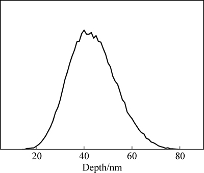
ͼ1 Gd����ע���Mg-Nd-Sr-Zr�Ͻ���GdԪ�طֲ�
Fig. 1 Gd element distribution in Mg-Nd-Sr-Zr alloy implanted by Gd ions
����SRIM 2008������Gd����ע��Mg-Nd-Sr-Zr�Ͻ���ע��Ԫ�طֲ�����ģ�⣬������ͼ1��ʾ����ͼ1���Կ�����ע��Ԫ����þ�Ͻ�����гʸ�˹�ֲ�������������Լ80 nm���ھ������Լ40 nm����ȴ���GdԪ��Ũ�ȴﵽ��ߡ�������̬�ȶ�������[22]��֪��ע��������ı�ע��Ԫ���ںϽ��е����(�����)�����Ũ��λ�ã���ˣ���ģ������֪��3�ּ�����Gd���Բ�ĺ��ԼΪ80 nm�����ھ����Լ40 nm����ȴ�GdԪ��Ũ����ߡ�
2.2 ����֯����
ͼ2��ʾΪMg-Nd-Sr-Zr�Ͻ���ܴ�����Ĺ�ѧ����֯����ͼ2���Կ������Ͻ���֯��Ҫ�ɦ�-Mg�������ڶ����Լ�������ɫϸ������ɡ�����Sr��Mg�й��ܶ����ޣ���ˣ�Sr���ܳ������Mg�����ڣ��Եڶ�����ʽ�����ھ��紦��ͨ��XRD���������ڶ���ΪMg41Nd5��Mg17Sr2[23]�����⣬ǰ�ڹ����ѷ����ڦ�-Mg�����ڹ۲쵽�Ŀ���״����״��ɫϸ����Ϊ��Zr������[19]��
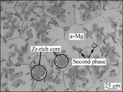
ͼ2 ����̬Mg-Nd-Sr-Zr�Ͻ�����֯
Fig. 2 Microstructure of solution treated Mg-Nd-Sr-Zr alloy
2.3 XRD��XPS����
ͼ3��ʾΪþ�Ͻ�ע��Gdǰ���XRD�ס���ͼ3���Կ���������þ�Ͻ��XRD����Ҫ�Ե��͵Ħ�-Mg�����Ϊ��������������Mg41Nd5��Mg17Sr2�ڶ�������塣��Gd����ע���þ�Ͻ���������г��˦�-Mg�͵ڶ���������⣬������Gd2O3��Nd2O3��Gd������壬������ע����������ӣ����������ǿ�ȳ���ǿ���ơ����⣬��ͼ�оֲ������Ŵ���Է��֣�����ע���-Mg����巢��ƫ�ƣ���˵���ֲ��������仯����ԭ����������Ӻ��þ�Ͻ����ʱ������ԭ����ײ�����Ա��Σ������侧���ڲ�����Ӧ��[24-25]��
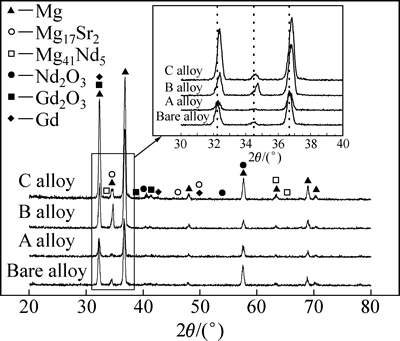
ͼ3 þ�Ͻ�ע��Gdǰ���XRD��
Fig. 3 XRD patterns of magnesium alloy before and after Gd implantation
����ע���ĺ�Ƚϱ�(Լ80 nm)����ʱXRD�ɼ���ע��������ź����ޣ���ˣ���ʵ���в���XPS��ע���(B�Ͻ�)�����˽�һ��������Ϊ����ź��ռ��������Ȳ�������ӽ�ע��þ�Ͻ�����GdŨ����ߴ�(�������Լ40 nm����ȴ�)���ɴ˵õ�XPS����ͼ4��ʾ����ͼ4���Կ�����ע�������Mg��Nd��Gd��Sr��Zr��O��C��Ԫ�س��֡�OԪ��Դ��������в���Ŀ�����������CԪ�ؿ�����Դ����ϴ���Ҵ���Һ���������ʣ�Sr��ZrԪ����þ�Ͻ�����Ԫ�أ���ע����еĺ������١�ͼ5��ʾΪMg 1s��Gd 4d��Nd 3d��XPS�ס���ͼ5(a)�п��Կ�����Mg 1s��������Ϊ977.50 eV�ķ�λ����ӦMgO��ͼ5(b)��ʾ��Gd 4d������˫��ṹ�������ֱ�Ϊ142.7��148.1 eV����ӦGd 4d5/2��Gd 4d3/2���ֱ���Gd��Gd2O3��λֵ���Ǻ�[26]��Nd 3dͬ������977.50��983.10 eV������λ(��ͼ5(c))����Nd2O3��λֵ��Ӧ[27]��������XPS���Խ����֪��Gd����ע��Mg-Nd-Sr-Zr�Ͻ��������γ���Gd2O3��Nd2O3������MgO������̬Gd��ɵĻ�ϲ㣬�ò��Խ����XRD���Խ�������Ǻϡ�
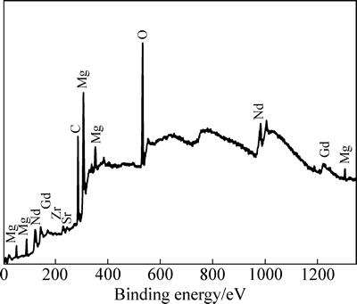
ͼ4 ע����XPSȫ��ͼ
Fig. 4 XPS survey spectra of implanted layer
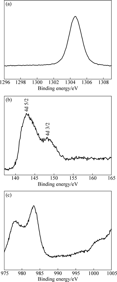
ͼ5 ע�����Mg 1s�塢Gd 4d�弰Nd 3d��ĸ߷ֱ���ͼ
Fig. 5 High-resolution XPS spectra of Mg 1s (a), Gd 4d (b) and Nd 3d (c) peakes of implanted layer
2.4 ���︯ʴ��Ϊ����
ͼ6��ʾΪ����þ�Ͻ��ע��þ�Ͻ���SBF��Һ�н���1 h�ļ������ߡ���ͼ6��֪��Gd����ע���þ�Ͻ�ļ�������������λ�����ƶ���˵��Gd����ע����ǿ��þ�Ͻ���SBF�е���ʴ���ܡ��ɼ���������ϵõ��ĸ�ʴ��λ�븯ʴ�����ܶ����1��ʾ����������ݿ�֪����ע����������ӣ�þ�Ͻ�ĸ�ʴ��λ�������ߣ���ʴ�����ܶ��Ƚ��ͺ���������ʴ�����ܶ��븯ʴ����������أ��Ҹ�ʴ�����ܶ�ԽС����ʴ����Խ������ע�����Ϊ5��1016 cm-2ʱ��ע��þ�Ͻ�(��B�Ͻ�)�ĸ�ʴ�����ܶ���С��Ϊ6.7��A/cm2�����ڻ���������������˵��B�Ͻ���SBF�еĸ�ʴ������������ע������������ӣ�ע��þ�Ͻ�ĸ�ʴ�����ֿ�ʼ�ӿ죬˵����������Gd����ע�������þ�Ͻ���ʴ������ǿ�̶ȡ�
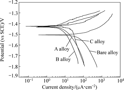
ͼ6 ����þ�Ͻ��ע��þ�Ͻ���SBF��Һ�еļ�������
Fig. 6 Polarization curves of bare and implanted magnesium alloys in SBF
��1 �Ͻ�ļ���������Ͻ��
Table 1 Fitted results of polarization curves of alloys

ͼ7��ʾΪ����þ�Ͻ���ע��þ�Ͻ���SBF�����������������ʱ��ı仯���ߡ���ͼ7�ɼ���3��ע��þ�Ͻ���ֳ���ͬ���������ƣ����Ž���ʱ���ӳ������������������ߺ��������ȶ�������A�Ͻ�����������٣�B�Ͻ�Ĵ�֮��C�Ͻ����ࡣ������þ�Ͻ���������������Ž���ʱ���ӳ��������ߣ��������������Զ�������3��ע��Ͻ�����������������ȣ�A��B��C 3�ֺϽ�ĸ�ʴ���ʷֱ�81%��79%��70%����˵��Gd����ע������Ч����þ�Ͻ���SBF�еĸ�ʴ���ʣ���A�Ͻ���SBF�еĸ�ʴ������С����ע��������ߣ�þ�Ͻ���ʴ����ǿ�̶Ƚ��͡����ݳ��ڣ��Ͻ�����������٣�����Դ��������γ��˱���Ĥ����һ���̶��������˺Ͻ�ĸ�ʴ��
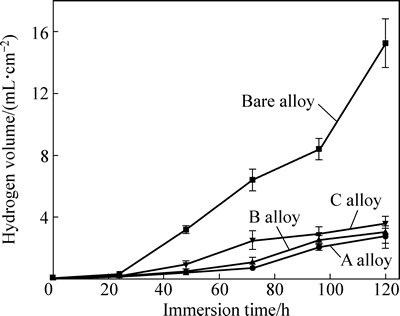
ͼ7 ����þ�Ͻ���ע��þ�Ͻ���SBF�е���������
Fig. 7 Hydrogen evolution curves of bare and implanted alloys in SBF
ͨ���绯ѧʵ�鼰����ʵ�������֪����Gd����ע���þ�Ͻ���SBF�еĸ�ʴ�������Լ������ҹ�����������ע��������þ�Ͻ�ʴ���������á����绯ѧʵ��������ʵ�鷴ӳ���Ľ�����в�ͬ���绯ѧʵ����ʾB�Ͻ���SBF�еĸ�ʴ������С��������ʵ�����A�Ͻ���SBF�и�ʴ�������������ֲ��Է����Ĺ��������йأ��绯ѧʵ����Ե���˲ʱ����������ȶ�����������ʵ���ܹ���ʵ��ӳ����ʴ������ʱ��仯�ı���[28]����ˣ�������þ�Ͻ�Ľ�����Ϊ���ԣ�����ʵ��IJ��Խ���ȵ绯ѧʵ����Խ�����ɿ���
ͼ8��ʾΪ����þ�Ͻ���ע��þ�Ͻ�(A�Ͻ�)��SBF�н���120 h��ĸ�ʴ��òͼ����ͼ8���Կ���������þ�Ͻ���渽��һ����ĸ�ʴ����Ͻ������ȫ��ʴ����ע��þ�Ͻ������ȷֲ��Ž��ٵĸ�ʴ������Դ���δ��ʴ�������һ��˵������ע���þ�Ͻ�ĸ�ʴ���ʽ��͡�
þ�Ͻ���SBF�з�����ʴ��ҪԴ����Һ��Cl-����ʴ����ˮ��Һ�У�þ�Ͻ��е�þԪ����ˮ��Ӧ���ڱ����������ɵ�Mg(OH)2����һ���̶����谭�˸�ʴ��Һ���к����ӵ���ʴ����ˣ��ڸ�ʴ���ڣ�þ�Ͻ�ʴ���ʻ�����Ȼ�������Ÿ�ʴʱ����ӳ�����Һ�е�Cl-����Mg(OH)2�У�����ת��Ϊ������MgCl2����ɺϽ����Mg�ܽ⣬�γɸ�ʴ[29-30]��
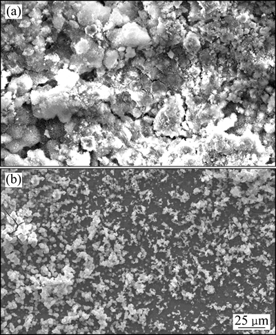
ͼ8 ����þ�Ͻ���ע��þ�Ͻ���SBF�н���120 h��ĸ�ʴ��ò
Fig. 8 Corrosion morphologies of bare (a) and implanted (b) alloys immersed in SBF for 120 h
���ڻ���þ�Ͻ���û�������ֲ�����ʴ�Եڶ���(��ͼ2)����Cl-��ȱ���㹻�ĵ�������[31]��ͬʱ�������ĵڶ����Zr�����������þ֮���γ���ż���Ӷ�����Cl-��þ�Ͻ������ʴ[31-32]����ˣ�������Mg�ܽ�ӿ졣��þ�Ͻ�Gd����ע���þ�Ͻ����ʴ����ǿ������������γɵĸ��Բ��йء����ڸ��Բ���Gd2O3[33]��Nd2O3[34]������Һ���ȶ����ܹ���Ч�谭Cl-���ڲ�þ�Ͻ���崫�ݣ��Ӷ�������Cl-��������Ӵ��ʣ������˻�����Mg�ܽ��ٶȡ����⣬ʵ���л����֣���ע���������(1��1017 cm-2)ʱ������ע��ĸ���Ч���½���WANG��[35-36]Ҳ�������Ƶ�������������ע�����Ӷ�þ�Ͻ�����������ã��������Բ�ı�����������ע�����Խ�Ը��Բ�����Խ���أ��䱣��Ч���½�[35-36]���ɱ�ʵ���֪������ע���þ�Ͻ���澧�����˱仯����������������Ӻ����ɾ����ڲ�����Ӧ�����Ӷ���������ע�����Ч����������ԭ����Ҫ���н�һ�����о���
3 ����
1) ��SRIM 2008����ģ���֪��GdԪ����þ�Ͻ�����гʸ�˹�ֲ���������Լ80 nm�����ھ������Լ40 nm��ȴ���GdԪ��Ũ�ȴﵽ��ߡ�
2) ����̬Mg-Nd-Sr-Zr�Ͻ�Gd����ע�����������γ�����Gd2O3��Nd2O3������MgO������̬Gd��ɵĸ��Բ㡣
3) ��Gd����ע���þ�Ͻ���SBF�еĸ�ʴ�������Լ�����������ע��������ߣ�������þ�Ͻ���ʴ����ǿ�̶ȡ�����ʵ�������ɿ���ӳ��þ�Ͻ���SBF�е����︯ʴ��Ϊ����ע�����Ϊ2.5��1016 cm-2ʱ��ע��þ�Ͻ�ĸ�ʴ������ͣ��������ȣ��丯ʴ���ʽ���81%��
REFERENCES
[1] WU G S, IBRAHIM J M, CHU P K. Surface design of biodegradable magnesium alloys-a review[J]. Surface and Coatings Technology, 2013, 233: 2-12.
[2] ������, Ѧ�Ǿ�, ������, ���Դ�, �� ǿ. Mg-(4-x)Nd-xGd-Sr-Zn-Zr ����þ�Ͻ����֯����ѧ��ʴ����[J]. ����ѧ��, 2014, 50(8): 979-988.
ZHANG Xiao-bo, XUE Ya-jun, WANG Zhang-zhong, HE Xian-cong, WANG Qiang. Microstructure, mechanical and corrosion properties of Mg-(4-x)Nd-xGd-Sr-Zn-Zr biomagnesium alloys[J]. Acta Metallurgica Sinica, 2014, 50(8): 979-988.
[3] WAN Y Z, XIONG G Y, LUO H L. Influence of zinc ion implantation on surface nanomechanical performance and corrosion resistance of biomedical magnesium-calcium alloys[J]. Applied Surface Science, 2008, 254(17): 5514-5516.
[4] ������, Ԭ����, ������. ����þ�Ͻ�Mg-Nd-Zn-Zr�����︯ʴ����[J]. �й���ɫ����ѧ��, 2013, 23(4): 905-911.
ZHANG Xiao-bo, YUAN Guang-yin, WANG Zhang-zhong. Biocorrosion properties of as-cast Mg-Nd-Zn-Zr magnesium alloy[J]. The Chinese Journal of Nonferrous Metals, 2013, 23(4): 905-911.
[5] WU G S, ZHANG X M, ZHAO Y, IBRAHIM J M, YUAN G Y, CHU P K. Plasma modified Mg-Nd-Zn-Zr alloy with enhanced surface corrosion resistance[J]. Corrosion Science, 2014, 78: 121-129.
[6] YAN Ting-ting, TAN Li-li, XIONG Dang-sheng, LIU Xin-jie, ZHANG Bing-chun, YANG Ke. Fluoride treatment and in vitro corrosion behavior of an AZ31B Mg alloy[J]. Materials Science and Engineering A, 2010, 30(5): 740-748.
[7] ������, ������, �� ǿ, ��־��, ������. þ�Ͻ�������þ��Ĥ�γɻ��Ƽ������︯ʴ����[J]. �й���ɫ����ѧ��, 2014, 24(5): 1285-1292.
FANG Xin-xian, ZHANG Xiao-bo, WANG Qiang, BA Zhi-xin, WANG Zhang-zhong. Formation mechanism and bio-corrosion properties of NaMgF3 film on surface of magnesium alloy[J].The Chinese Journal of Nonferrous Metals, 2014, 24(5): 1285-1292.
[8] SANTHANAKRISHNAN S, HO Y H, DAHOTRE N B. Laser coating of hydroxyapatite on Mg for enhanced physiological corrosion resistance and biodegradability[J]. Materials Technology, 2012, 27: 273-277.
[9] TALTAVULL C, TORRES B, LOPEZ A J, RODRIGO P, OTERO E, ATRENS A, RAMS J. Corrosion behaviour of laser surface melted magnesium alloy AZ91D[J]. Materials Design, 2014, 57: 40-50.
[10] LIN X, YANG X M, TAN L L, LI M, WANG X, ZHANG Y, YANG K, HU Z Q, QIU J H. In vitro degradation and biocompatibility of a strontium-containing micro-arc oxidation coating on the biodegradable ZK60 magnesium alloy[J]. Applied Surface Science, 2014, 288: 718-726.
[11] YANG X M, LI M, LIN X, TAN L L, LAN G B, LI L H, YIN Q S, XIA H, ZHANG Y, YANG K. Enhanced in vitro biocompatibility/bioactivity of biodegradable Mg-Zn-Zr alloy by micro-arc oxidation coating contained Mg2SiO4[J]. Surface and Coatings Technology, 2013, 233: 65-73.
[12] ZHAO Y, JAMESH M I, LI W K, WU G S, WANG C X, ZHANG Y F, YEUNG K W K, CHU P K. Enhanced antimicrobial properties, cytocompatibility, and corrosion resistance of plasma-modified biodegradable magnesium alloys[J]. Acta Biomaterialia, 2014, 10: 544-556.
[13] WU G S, XU R Z, FENG K. Retardation of surface corrosion of biodegradable magnesium- based materials by aluminum ion implantation[J]. Applied Surface Science, 2012, 258(15): 7651-7657.
[14] WU G S, GONG L, FENG K. Rapid degradation of biomedical magnesium induced zinc ion implantation[J]. Materials Letters, 2011, 65(4): 661-663.
[15] WANG Z Z, TAO X W, ZHANG X B, BA Z X, WANG Q. Corrosion behaviour of Nd ion implanted Mg-Gd-Zn-Zr alloy in simulated body fluid[J]. Materials Technology, 2015, 30(6): 321-325.
[16] JIN W H, WU G S, FENG H Q, WANG W H, ZHANG W M, CHU P K. Improvement of corrosion resistance and biocompatibility of rare-earth WE43 magnesium alloy by neodymium self-ion implantation[J]. Corrosion Science, 2015, 94: 142-155.
[17] WANG W J, ZHANG X L, WU G S, WANG C X, CHU P K. Praseodymium-surface- modified magnesium alloy: Retardation of corrosion in artificial hands weat[J]. Materials Letters, 2016, 163: 85-89.
[18] GEROLD B. Implant with a base body of a biocorrodible magnesium alloy[P]. US 8268235 B2, 2012-09-18.
[19] ZHANG X B, WU Y J, XUE Y J, WANG Z Z, YANG L. Biocorrosion behavior and cytotoxicity of a Mg-Gd-Zn-Zr alloy with long period stacking ordered structure[J]. Materials Letters, 2012, 86: 42-45.
[20] ZHANG Xiao-bo, BA Zhi-xin, WANG Zhang-zhong, XUE Ya-jun, WANG Qiang. Microstructure and biocorrosion behaviors of solution treated and as-extruded Mg-2.2Nd-xSr-0.3Zr alloys[J]. Transactions of Nonferrous Metals Society of China, 2014, 24: 3797-3803.
[21] TAO X W, WANG Z Z, ZHANG X B, BA Z X, WANG Y M. Nanomechanical and corrosion properties of ZK60 magnesium alloy improved by Gd ion implantation[J]. Surface Review and Letters, 2014, 21(6): 1450085- (1-6).
[22] ��ͨ��, ��褹�. ����ע������Ż�����[M]. ����: ұ��ҵ������, 1993.
ZHANG Tong-he, WU Yu-guang. Optimizing pressing of ion implantation[M]. Beijing: Metallurgical Industry Press, 1993.
[23] ZHANG X B, HE X C, XUE Y J, WANG Z Z, WANG Q. Effects of Sr on microstructure and corrosion resistance in simulated body fluid of as cast Mg-Nd-Zr magnesium alloys[J]. Corrosion Engineering, Science and Technology, 2014, 49(5): 345-351.
[24] LIU B J, DENG B, TAO Y. Influence of niobiumion implantation on the microstructure, mechanical and tribological properties of TiAlN/CrN nano-multilayer coatings[J]. Surface and Coatings Technology, 2014, 240: 405-412.
[25] LIU H X, XU Q, JIANG Y H, WANG C Q, ZHANG X W. Corrosion resistance and mechanical property of AZ31 magnesium alloy by N/Ti duplex ion implantation[J]. Surface and Coatings Technology, 2013, 228: 538-543.
[26] RAISER D, DEVILLE J P. Study of XPS photoemission of some gadolinium compounds[J]. Journal of Electron Spectroscopy and Related Phenomena, 1991, 57(1): 91-97.
[27] UWAMINO Y, ISHIZUKA T, YAMATERA H. X-ray photoelectron spectroscopy of rare-earth compounds[J]. Journal of Electron Spectroscopy and Related Phenomena, 2012, 34(1): 67-78.
[28] KIRKLAN N T, BIRBILIS N, STAIGER M P. Assessing the corrosion of biodegradable magnesium implant: a critical review of current methodologies and their limitations[J]. Acta Biomaterialia, 2012, 8: 925-936.
[29] ARDELEAN H, SEYEUX A, ZANNA S, PRIMA F, FRATEUR I. MARCUS P. Corrosion processes of Mg-Y-Nd-Zr alloys in Na2SO4 electrolyte[J]. Corrosion Science, 2013, 73: 196-207.
[30] ZHAO Y Z, WU G S, LIU Q, WU J, XU R,YEUNG K W K, CHU P K. Improved surface corrosion resistance of WE43 magnesium alloy by dual titanium and oxygen ion implantation[J]. Thin Solid Films, 2013, 529: 407-411.
[31] �� ��, ������, ���ϴ�, �����, ������, ������. �ڶ���������Mg-Gd-Y-Nd-Zr�Ͻ�ֲ���ʴ�е����û���[J]. �й���ɫ����ѧ��, 2013, 23(1): 15-21.
LIU Jun, CHEN Ming-an, MA Cong-cong, HUANG Yu-di, ZHANG Xin-ming, DENG Yun-lai. Effect of second phase particles on localized corrosion of Mg-Gd-Y-Nd-Zr alloy[J]. The Chinese Journal of Nonferrous Metals, 2013, 23(1): 15-21.
[32] SUN M, WU G H, WANG W, DING W J. Effect of Zr on the microstructure, mechanical properties and corrosion resistance of Mg-10Gd-3Y magnesium alloy[J]. Materials Science and Engineering A, 2009, 523: 145-151.
[33] UNE K, KASHIBE S, NOGITA K. Corrosion behavior of unirradiated oxide fuel pellets in high temperature water[J]. Journal of Nuclear Materials, 1995, 227(1/2): 32-39.
[34] CHEVALIERA S, BONNETB G, LARPINA J P. Metal-organic chemical vapor deposition of Cr2O3 and Nd2O3 coatings. Oxide growth kinetics and characterization[J]. Applied Surface Science, 2000, 167: 125-133.
[35] WANG X M, ZENG X Q, YAO S S, WU G S, LAI Y J. The corrosion behavior of Ce-implanted magnesium alloys[J]. Materials Characterization, 2008, 59(5): 618-623.
[36] WANG X M, ZENG X Q, WU G S, YAO S S. Yttrium ion implantation on the surface properties of magnesium[J]. Applied Surface Science, 2006, 253: 2437-2442.
Effect of Gd ion implantation on biocorrosion behavior of solution treated Mg-Nd-Sr-Zr alloy
TAO Xue-wei1, 2, WANG Zhang-zhong1, 2, ZHANG Xiao-bo1, 2, BA Zhi-xin1, 2, DONG Qiang-sheng1, 2
(1. School of Materials Science and Engineering, Nanjing Institute of Technology, Nanjing 211167, China;
2 Jiangsu Key Laboratory of Advanced Structural Materials and Application Technology,
Nanjing Institute of Technology, Nanjing 211167, China)
Abstract: Gadolinium (Gd) ion implantation was carried out to modify the solution-treated Mg-Nd-Sr-Zr alloy. The SRIM 2008 software was used to simulate the Gd ion implantation. The microstructure of alloy was observed by optical microscopy (OM). The phases were analyzed by X-ray diffractometry (XRD), and the chemical composition and element states of modified layer were characterized by X-ray photoelectron spectroscopy (XPS). The biocorrosion behavior of the bare and Gd-implanted alloys in simulated body fluids (SBF) was evaluated by electrochemical tests and hydrogen evolution tests. The results show that the biocorrosion resistance of solution treated Mg-Nd-Sr-Zr alloy is enhanced after Gd ion implantation. When the implantation dose is 2.5��1016 cm-2, the implanted alloy has the optimal anti-biocorrosion performance.
Key words: magnesium alloy; Gd; ion implantation; biocorrosion behavior
Foundation item: Project(51301089) supported by the National Natural Science Foundation of China; Project supported by Qing Lan Project of Jiangsu Province, China
Received date: 2015-06-29; Accepted date: 2015-12-02
Corresponding author: WANG Zhang-zhong; Tel: +86-025-86118275; E-mail: zzww@njit.edu.cn
(�༭ ������)
������Ŀ��������Ȼ��ѧ����������Ŀ(51301089)������ʡ���������̡�������Ŀ
�ո����ڣ�2015-06-29�������ڣ�2015-12-02
ͨ�����ߣ������ң����ڣ��绰��025-86118275��E-mail��zzww@njit.edu.cn
ժ Ҫ���Թ���̬Mg-Nd-Sr-Zr�Ͻ��������(Gd)����ע����Դ���������SRIM 2008������Gd����ע����̽�����ģ����������ù�ѧ����(OM)�۲���þ�Ͻ������֯��������X ����������(XRD)�������Բ��е�������ɣ�ͬʱ���X���߹��������(XPS)�������Բ��л�ѧ�ɷ���Ԫ�ؼ�̬�����õ绯ѧʵ�鼰����ʵ�������˻���þ�Ͻ���ע��þ�Ͻ���ģ����Һ(SBF)�е����︯ʴ��Ϊ�����������Gd����ע����������߹���̬Mg-Nd-Sr-Zr�Ͻ���SBF�������︯ʴ���ܣ��ҵ�ע�����Ϊ2.5��1016 cm-2ʱ��ע��þ�Ͻ�������︯ʴ������ѡ�
