Preparation of poly-L-lactide/bioactive glass composite and evaluation of cytotoxicity in vitro
来源期刊:中国有色金属学报(英文版)2008年第5期
论文作者:周智华 阮建明 邹俭鹏 周忠诚 陈良龙
文章页码:1151 - 1151
Key words:poly-L-lactide; bioactive glass; composite; cytotoxicity; biocompatibility; in vitro
Abstract: Bioactive and bioresorbable composite materials were fabricated from poly-L-lactide and bioactive glass (average particle size 6.8 μm) by a solvent evaporation technique. Cellular cultivation in vitro and MTT assay were conducted for evaluating the influence on morphology, growth and proliferation of cultured fibroblasts. The results of cytotoxicity testing show that cells cultured in extracts of PLLA/BG and on the surface of composites demonstrate normal growth and proliferation. The bioactive glass in PLLA composite facilitates both adhesion and proliferation of rat fibroblasts on PLLA/bioactive glass composite film.
基金信息:the National Natural Science Foundation of China

ZHOU Zhi-hua(周智华)1, 2, RUAN Jian-ming(阮建明)1, ZOU Jian-peng(邹俭鹏)1,
ZHOU Zhong-cheng(周忠诚)1, CHEN Liang-long(陈良龙)3
1. State Key Laboratory of Powder Metallurgy, Central South University, Changsha 410083, China;
2. College of Chemistry and Chemical Engineering, Hunan University of Science and Technology,
Xiangtan 411201, China;
3. Department of Orthopedics, the Second Xiangya Hospital, Central South University, Changsha 410011, China
Received 29 October 2007; accepted 29 December 2007
Abstract: Bioactive and bioresorbable composite materials were fabricated from poly-L-lactide and bioactive glass (average particle size 6.8 μm) by a solvent evaporation technique. Cellular cultivation in vitro and MTT assay were conducted for evaluating the influence on morphology, growth and proliferation of cultured fibroblasts. The results of cytotoxicity testing show that cells cultured in extracts of PLLA/BG and on the surface of composites demonstrate normal growth and proliferation. The bioactive glass in PLLA composite facilitates both adhesion and proliferation of rat fibroblasts on PLLA/bioactive glass composite film.
Key words: poly-L-lactide; bioactive glass; composite; cytotoxicity; biocompatibility; in vitro
1 Introduction
Being environmentally friendly over synthetic polymers, poly-L-lactide(PLLA) is a biodegradable and biocompatible semi-crystalline polymer. It may be used as controlled-release devices, absorbable sutures, implants in orthopaedics, absorbable fibres and disposable degradable plastics. Many investigations have been published on the degradation of PLLA both in vitro and in vivo[1-2]. PLLA can react with something as a carrier supporting tissue for a given length of time prior to be gradually biodegraded. A major drawback of this material is its release of acidic degradation products which may lead to inflammatory responses[3]. Another limitation is its lack of bioactivity. In the case of bone tissue engineering, it does not allow bone apposition or bonding on polymer surface[4].
Bioactive glass(BG) was developed by Hench in early 1970s, and has remained as the choice of bone- bioactive material from then[5]. Its bioactivity or osteointegration potential is directly related to the formation of a surface calcium phosphate(Ca-P) layer[6]. The biocompatibility, osteoconductivity, and osteoinduc- tivity of BG have been well documented[7]. However, the direct application of BG in load-bearing applications has been limited due to its brittle and poor tensile and torsional properties.
The PLLA/BG composite was designed to integrate the advantages of parent phases and to minimize those well-known limitations associated with each component. A significant advantage of PLLA/BG over PLLA is its osteointegration potential or the ability to form a Ca-P surface layer in vitro[8]. Osteointegration is a critical factor in facilitating the chemical fixation of a biomaterial to bone tissue. The second advantage of the composite is that the addition of BG to PLLA matrix results in a structure with higher compressive modulus. A successful internal fixation material must exhibit mechanical properties similar to those of the tissue to be replaced[9]. Therefore, the PLLA/BG composite would render greater functionality in vivo compared with PLLA. Moreover, the combination of the two phases serves to neutralize both the acidic byproducts produced during polymer degradation[10-11] and the alkalinity due to the formation of calcium phosphate layer.
Toxic organic solvent CH2Cl2 was used during the course of preparing PLLA/BG composite. Since most implant materials are implanted into human body in direct contact with human tissue, they are not allowed to be toxic or immunogenic, not to induce neoformation of the surrounding tissues and not to inhibit re- establishment of the normal function. Therefore, they are required to carry out toxicity and biocompatibility evaluation before being applied to the clinic. Both toxicity and biocompatibility evaluations have been proven to be valuable in vitro for testing and screening candidate biomaterials[12].
In this work, a solvent evaporation technique was used to process PLLA/BG composite film. The cytotoxicity of the composite in vitro was carried out by cellular cultivation and MTT assay.
2 Experimental
2.1 Preparation of PLLA/BG composite
Homemade L-lactide was used as a monomer for PLLA synthesis. Stannous octoate was used as an initiator without additional purification. Ampoules for polymerization were dried overnight in an oven at 400 ℃. Then, they were loaded with freshly recrystallized L-lactide and Sn-octoate toluene solution with ratios 1 000-12 000 of monomer to initiator. After that, they were sealed under high vacuum, and left in the oven at 140 ℃, so that the monomer could polymerize[13]. The polymerization time varied from 12 to 60 h. The polymer was dissolved in chloroform, precipitated in methanol and dried in vacuum. PLLA has obtained a viscosity-average relative molecular mass about 4.0×10⁵. Bioactive glass with an average particle size of 6.8 μm and a composition of 35CaO, 60SiO2, 5P2O5 (mole fraction, %) was also prepared by sol-gel method according to Ref.[14].
PLLA/bioactive glass composite was prepared by dissolving polymer in CH2Cl2 to produce a polymer mass with solvent volume ratio of 5% (w/v). The mixture was stirred overnight to obtain a homogeneous solution. Bioactive glass powder (10%, mass fraction) was added into polymer solution. The mixture was transferred into a flask and sonicated for 60 min to improve the dispersion of the bioactive glass particles. After homogenization, the mixture was cast onto a flat glass plate to form a membrane. The membrane was then dried in a vacuum oven at 40 ℃.
2.2 Preparation of PLLA/BG extracts
PLLA/BG composites were placed in tissue culture flasks with RPMI-1640 medium containing 10% fetal calf serum (FCS) of 0.1 cm2/mL and incubated in a cell incubator for 48 h (100% relative humidity and 5% CO2). Positive controls contained medium of 0.64% phenol and negative controls contained only RPMI-1640 medium with 10% fetal calf serum (FCS).
2.3 Cell morphology cultured in extracts
Cell lines rat fibroblasts were used for proliferation study. Rat fibroblasts were routinely grown and maintained in RPMI-1640 medium containing 10% fetal calf serum. Three groups of cells were seeded in 24-well plates with cell seeding density of 1×10⁵ in a humidified incubator containing 5% CO2 at 37 ℃. On the third day of the experiment, the medium from each well was removed and replaced with 2 mL of the extract from one of the specimens (or with the positive or negative control medium). Six wells were prepared for each extracts, positive controls and negative controls. Cell morphology was observed under the reverse microscope (TE2000U, NIKON)
2.4 MTT measurement
MTT assay was based on the protocol described for the first time by MOSSMANN. The assay was optimized for the cell lines in the experiments. Three groups of cells were seeded in 96-well plates with cell seeding density of 1×10⁵ in a humidified incubator containing 5% CO2 at 37 ℃ for 24 h. Then, cells were incubated for 24 h in extracts of PLLA/BG, negative control medium and positive control medium containing 10% FCS and 0.64% phenol, respectively. At the end of the incubation, cells were incubated for 4 h with 10 ?mol/L of MTT. Washing with PBS was followed by the addition of DMSO (150 μL) and gently shaking for 10 min so that complete dissolution was achieved. The optical density (OD) was measured at 492 nm[15]. The relative growth rate(RGR) was calculated according to the following equation:
RGR=ODsamples/ODnegative control×100% (1)
2.5 Cell morphology on surface of PLLA/bioactive glass composite and PLLA
Cell attachment and cell morphology on PLLA/bioactive glass and PLLA at different time interval were studied. Cover-slides coated with PLLA/bioactive glass and PLLA were placed into 24-well plates (costar), washed three times with PBS. 3 mL medium was added to the wells to prevent the cover slide from floating during cell seeding. 1.0×10⁵ cells (in 1 mL of medium) were then placed in each well and the plates were incubated at 37 ℃ with 5% CO2 for 7 d. The cover-slides were washed three times with PBS and fixed with 3% glutaraldehyde in PBS at room temperature for 30 min. After thorough washing, the cells were dyed with one drop of Giemsa stain (SIGMA) for 30 min, followed by washing with distilled water and drying in air. Cell attachment and cell morphology were observed under reverse microscope (TE2000U, NIKON) and scanning electron microscope (KYKY-2800).
2.6 Statistical analysis
All measurements were collected and expressed as means ± standard deviations.Single factor analysis of variance was employed to assess the statistical significance of results for all biological experiments.
3 Results and discussion
3.1 Characterization of PLLA/BG composite
The surface morphology of the composite is shown in Fig.1. Bioactive glass prepared has an average size of 6.8 μm with agglomeration. SEM micrograph provides the evidence of a small percentage of large glass particles (>10 μm). The bioactive glass particles, small or large, are angular in shape. Bioactive glass particles are distributed evenly in the composite nevertheless the polymer covers the bioactive glass particles on the surface.

Fig.1 SEM micrograph of PLLA/bioactive glass
The XRD pattern of the surface of composite is shown in Fig.2. This is a low crystalline polymeric composite with two peaks at 16.63? and 18.86? corresponding to (200) and (203) reflection of PLLA, respectively.
3.2 Morphology of fibroblast cells cultured in extracts
In biomedical applications, it is necessary to investigate the biological behaviors of the PLLA/bioactive composites. The composites prepared in this study and control samples were subjected to the biocompatibility test using primary cultures of rat fibroblast cells as the model system. It has been well known that cell adhesion and proliferation is an important cellular process because it directly influences the proliferation of cells and formation of bone tissue.

Fig.2 XRD pattern of PLLA/bioactive glass composite
Fig.3 presents the morphology of fibroblast cells cultured in different media. Rat fibroblasts show higher proliferation when being cultured with ionic products from PLLA/BG composite materials than with control medium. Cells cultured in both extracts of PLLA/BG and negative control medium attach to the culture plate on the third day, indicating that fibroblasts have strong generation abilities with some cells in mitoses. However, cells cultured in positive control medium goes dead after three days. The cell culture results indicate that the extracts from PLLA/BG are non-cytotoxic and can enhance cellular activity.

Fig.3 Morphologies of cultured fibroblasts: (a) PLLA/BG group, 12 h; (b) PLLA/BG group, 3 d; (c) Negative control group, 3 d; (d) Positive control group, 3 d
3.3 MTT assay
MTT is a biochemical test widely used to assess cytotoxicity by measuring cell viability and proliferation in a qualitative way. This biochemical test is based on the reduction of MTT (which is water-soluble salt and has a yellow tonality) by cell mitochondrial enzyme succinate dehydrogenase, yielding a purple-color salt insoluble in water[16]. The salt absorbs at a wavelength of 492 nm because only living cells have the capability to metabolize the MTT, which gives a measurement of the viable cells[17].
The results of toxicity of PLLA/BG by MTT assay are presented in Table 1. The RGR of cells cultured in the extracts of PLLA/BG is higher than that of negative control group. However, positive control group shows apparent toxicity. This indicates that PLLA/BG composite has no toxicity. Moreover, the existence of bioactive glass in composites also demonstrates cell compatibility improvement due to good biocompatibility of the bioactive glass.
Table 1 Results of cytotoxicity levels by MTT assay

3.4 Cell attachment and morphology on surface of PLLA/BG composite
As seen in Fig.4, SEM was used to examine the morphology of the cells attaching and spreading on the films of PLLA/BG and PLLA after 7 days, respectively. The micrograph (Fig.4(a)) shows that there are no fibroblast cells attaching to the surface of PLLA. As shown in Fig.4(a), PLLA has a micropore and microcrack structure. This can be explained by the evaporation of CH2Cl2, which causes the shrinkage of the polymer. On the surface of PLLA/BG film (Fig.4(b)), fibroblast cells start to spread out and are very closely attached to the surface. The bulges of the main bodies of the fibroblast cells are still apparent and the spreading cells are readily distinguishable from the substrate.
As shown in Fig.4, the presence of bioactive glass particles in PLLA may have positive biological effects. The PLLA/bioactive glass composites also demonstrate improved cell compatibility due to the good biocompatibility of the bioactive glass particles and the more uniform distribution of the glass particles on the film surface. The loss of the bioactive glass particles in contact with the culture medium results in a coarse surface for the cell adhesion and proliferation. In general, cell behavior and interaction with a bioactive material surface are dependent on properties such as topography, surface charge and chemistry. In addition, the bioactive glass particles disengaged from composite and exposed to body fluid might induce a microenvironment change, i.e., the alkalinization of the medium, which has a positive influence on cell metabolism.
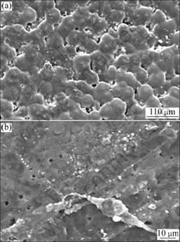
Fig.4 SEM micrographs of fibroblasts on surfaces of materials after 7 d: (a) PLLA; (b) PLLA/BG
When the PLLA/BG composite was immerged in body fluid, the water could easily permeate into the inner side of the composite, resulting in a rapid degradation from the outer side to the inner side and prompting loss of bioactive glass filler, owing to the relatively low inter- face adhesion between the particles and PLLA matrix, and several lacunas existed in the aggregated bioactive glass particles in composites. So, the PLLA/bioactive glass composite seemed to be more effective for the cell adhesion in the initial stage of cell culture.
Fig.5 presents the morphology of fibroblasts on the surface of PLLA/BG for different time by reverse microscope. Reverse microscope allows for the observation of the morphological changes of in vitro cell culture. After 12 h, cells appeared to show normal morphology with size ranging from 10 to 20 μm in diameter. The cells, assumed spindle-shaped, formed confluent layers on the composite surfaces after 3 weeks. Cells appeared to be normal in culture with PLLA/BG film, indicating that PLLA/BG composite is not cytotoxic. In addition, the smaller shape of cells around PLLA/BG composite film was similar to that of differentiated osteoblasts[18], suggesting that the cell differentiation process might be promoted in the presence of BG particles.

Fig.5 Micrographs of fibroblasts on surfaces of composites: (a) 12 h; (b) 3 weeks
4 Conclusions
1) A solvent evaporation technique was used to process PLLA/bioactive glass composites. Agglomerative bioactive glass granules were distributed homogeneously in the composite.
2) The cytotoxicity of PLLA/BG was evaluated by
rat fibroblasts culture in vitro and MTT test. The results of toxicity test show that the bioactive glass in the PLLA composite facilitates both adhesion and proliferation of rat fibroblast on the PLLA/BG composite film and extracting solution.
3) The PLLA/bioactive glass composite also demonstrates cell compatibility improvement due to good biocompatibility of the bioactive glass particles.
References
[1] ZHOU Z H, RUAN J M, ZOU J P, ZHOU Z C. The kinetics of melting crystallization of poly-L-lactide [J]. Polymer-Plastics Technology and Engineering, 2007, 46(9): 863-871.
[2] ROKKANEN P U, BOSTMAN O, HIRVENSALO E, MAKELA A, PARTIO E K, PATIALA H, VAINIONPAA S, VIHTONEN K, TORMALA P. Bioabsorbable fixation in orthopedic surgery and traumatology [J]. Biomaterials, 2000, 21(24): 2607-2613.
[3] AGRAWAL C M, RAY R B. Biodegradable polymeric scaffolds for musculoskeletal tissue engineering [J]. Biomed Mater Res, 2001, 55(2): 141-150.
[4] SCHLIEPHAKE H, NEUKAM F W, HUMACHER D. Enhancement of bone in-growth into a porous HA-matrix using a resorbable polylactice membrane [J]. J Oral Maxillofac Surg, 1994, 52(1): 57-63.
[5] ZHOU Z H, RUAN J M, ZOU J P, ZHOU Z C. Bioactivity of bioresorbable composite based on bioactive glass and poly-L-lactide [J]. Trans Nonferrous Met Soc China, 2007, 17(2): 394-399.
[6] VALLET-REGI M. Ceramics for medical applications [J]. Chem Soc Dalton Trans, 2001, 17(1): 97-108.
[7] ROETHER J A, BOCCACCINIB A R, HENCH L L, MAQUET V, GAUTIER S, JEROME R. Development and in vitro characterisation of novel bioresorbable and bioactive composite materials based on polylactide foams and bioglass? for tissue engineering applications [J]. Biomaterials, 2002, 23(18): 3871-3878.
[8] BONFIELD W. Design of bioactive ceramic-polymer composites [M]. Singapore: World Scientific, 1993: 299-303.
[9] BOCCACCINI A R, ROETHER J A, HENCH L L, MAQUET V, JEROOME R. Composites approach to tissue engineering [J]. Ceram Eng Sci Proc, 2002, 23: 805-816.
[10] NIEMELA T, KELLOMAKI M, TORMALA P. In vitro degradation of osteoconductive poly-L/DL-lactide/b-TCP composites [J]. Key Eng Mater, 2004, 54: 509-512.
[11] WANG M, HENCH L, BONFIELD W. Bioglass/high density polyethylene composite for soft tissue applications: Preparation and evaluation [J]. Biomed Mater Res, 1998, 42(4): 577-586.
[12] JONES J R, TSIGKOU O, COATES E E, STEVENS M M, POLAK J M, HENCH L L. Extracellular matrix formation and mineralization on a phosphate-free porous bioactive glass scaffold using primary human osteoblast (HOB) cells [J]. Biomaterials, 2007, 28(9): 1653-1663.
[13] ZHOU Z H, RUAN J M, ZOU J P, ZHOU Z C. Preparation of high viscosity average molecular weight poly-L-Lactide [J]. J Cent South Univ Technol, 2006, 13(6): 608-612.
[14] ZHOU Z H, RUAN J M, ZOU J P, ZHOU Z C. Synthesis and structural characterization of macroporous bioactive glass [J]. J Cent South Univ Technol, 2007, 14(3): 301-304.
[15] JOHSON H J. Biocompatibility test procedures for materials evaluation in vitro (I): Comparative test system sensitivity [J]. J Biomed Mater Res, 1983, 17(4): 571-579.
[16] PIETRZAK W S, EPPLEY B L. In vitro characteristics of a bioabsorbable suspension screw and suture system for endoscopic brow lift surgery [J]. J Craniofac Surg, 2007, 18(2): 429-436.
[17] BHANG S H, LIM J S, CHOI C Y, KWON Y K, HIM B S. The behavior of neural stem cells on biodegradable synthetic polymers [J]. J Biomater Sci Polym Ed, 2007, 18(2): 223-239.
[18] STEIN G S, LIAN J B, OWEN T S. Relationship of cell growth to the regulation of tissue-specific gene expression during osteoblast differentiation [J]. FASEB J, 1990, 4: 3111-3123.
Foundation item: Project(50771096) supported by the National Natural Science Foundation of China
Corresponding author: RUAN Jian-ming; Tel: +86-731-8876644; E-mail: jianming@mail.csu.edu.cn

