Precipitation Process of Copper-Containing Steel with Addition of Ni
Liang Mengfei Wang Haiyan Wang Chao Ren Huiping Gao Xueyun
School of Materials and Metallurgy,Inner Mongolia University of Science and Technology
Key Laboratory of Integrated Exploitation of Bayan Obo Multi-Metal Resource,Inner Mongolia University of Science and Technology
Abstract:
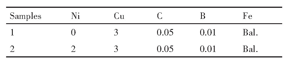
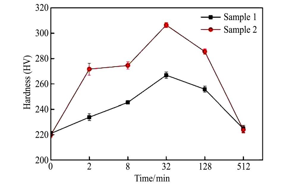
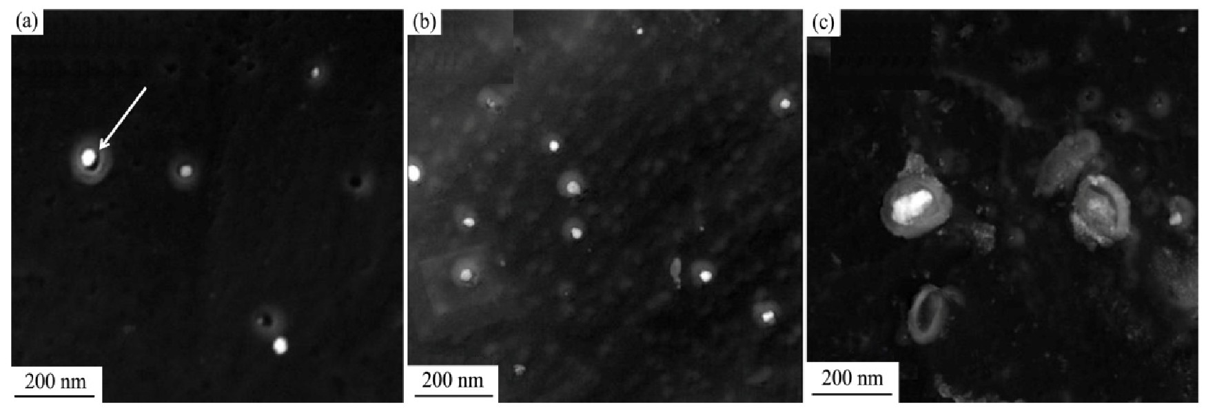
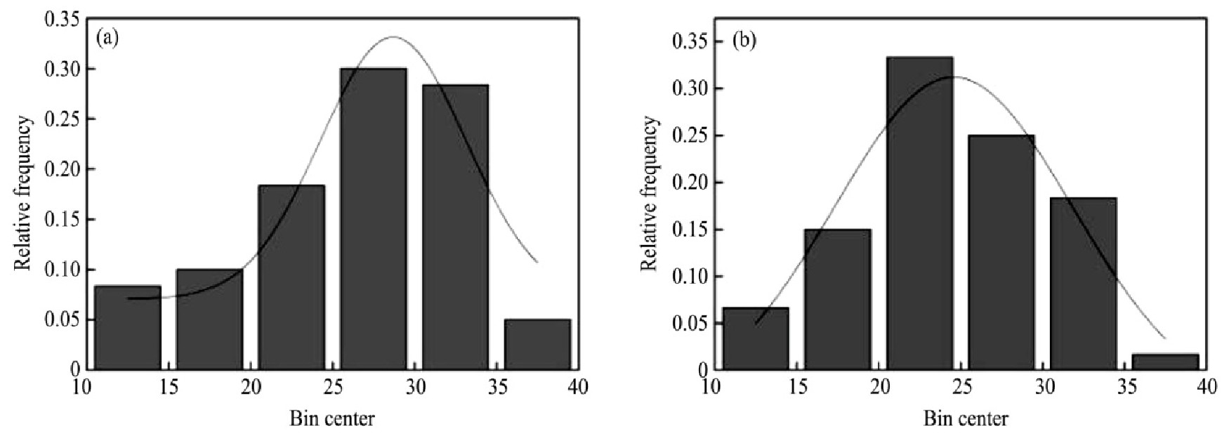
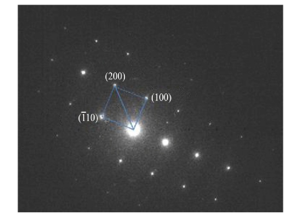
Copper is added to steel,and the precipitation strengthening effect produced by it makes steel materials have certain corro-sion resistance,high plasticity and high strength,as well as excellent welding performance and mechanical processing performance,which has a good development prospect.Domestic and foreign scholars design copper-containing steels with different compositions andformulate corresponding heat treatment processes,and conduct a lot of research on it according to the precipitation strengthening mech-anism of copper.Ni is an important element of high-strength alloy steel.It is mainly used to improve the rigidity of the matrix and thetoughness of the metal.Through its own solid solution strengthening effect,it can enhance the mechanical properties and improve theyield strength of the metal.Adding Ni element to Fe-Cu alloy,through Cu-rich particle precipitation experimental study,combinedwith theoretical analysis,were used to study the effect of Ni element addition on the Cu precipitation process of copper-containingsteel.Taking Fe-Cu alloys and Fe-Cu-Ni alloys before and after Ni addition as the research objects,the microstructure changes of FeCu alloys and Fe-Cu-Ni alloys during the aging process were observed with a metallurgical optical microscope(OM),the hardnesschange law of the two alloys during the isothermal aging process were analyzed with the help of hardness test,and transmission elec-tron microscopy(TEM)was used to observe the fine structure of the precipitated phase and diffraction analysis of the two alloys.Onthis basis,through the combination of experiment and theoretical analysis explored the influence of the addition of Ni on the micro-structure,hardness,precipitate morphology and size of copper-containing steel.The microscopic morphology and hardness testshowed that the addition of Ni improves the hardenability of the steel,thereby promoting the formation of quenched martensite,andthe strengthening effect of the alloy was significantly enhanced.The hardness was better than that of the experimental steel without theaddition of Ni.The reason might be that with the extension of the aging time,the internal structure of the experimental steel was con-stantly changing,the number of precipitated particles in the matrix was reduced,and the reduction of free energy made the nucleationrate slower,which led to the hardness after over-aging.Observation of the precipitated phase found that the addition of Ni in the cop-per-containing steel accelerated the nucleation process,increased the nucleation rate of the precipitated phase particles,promoted theprecipitation of the Cu-rich phase,and refined the size of the precipitated phase.The addition on the one hand changed the critical nu-cleation work of the Cu-rich phase precipitation,leading to changes in the size and number density of the precipitates.On the otherhand,it changed the distribution of elements in the Cu-rich phase precipitates,which could promote the Cu-rich phase precipitation,increase the nucleation rate and number density of the Cu-rich phase.During the growth of the Cu-rich phase,Ni would form a shellstructure at the Cu-rich phase/matrix interface,thereby reducing the coarsening rate of the Cu-rich phase,so that the Cu-rich phasesize remained small for a long time,thereby increasing the strength and hardness of the steel.Transmission electron diffraction analy-sis showed that during the growth process of the precipitated phase particles,bcc phase Cu particles were preferentially precipitated inthe matrix and coherent with the matrix belong to the metastable phase and would transform from bcc structure to fcc structure through dif-fusion and growth.During the aging process,the structure would evolve and the Cu-rich metastable phase of bcc structure would be grad-ually formed.With the extension of the aging time,the hardness of the experimental steel increased first and then decreased.The hard-ness of the experimental steel without Ni and the experimental steel with Ni reached the maximum value of HV 264.7 and HV 316.9 at 32 min.The addition of Ni made the alloy strengthening effect enhanced.In the quenched state,the microstructures of the experimental steelwithout Ni and the experimental steel with Ni were ferrite and lath martensite respectively.During the aging process,the microstructureof the experimental steel without Ni was still ferrite.The martensite structure in the experimental steel gradually transformed to ferrite.There were different morphologies such as spherical and ellipsoidal in the precipitation process of Cu particles.The addition of Ni ele-ment accelerated the nucleation process and refined the size of the precipitated phase.Cu diffused during the aging process,and the addi-tion of Ni increased the nucleation rate of the precipitated phase,promoted the precipitation of the Cu-rich phase,and formed a Cu-richmetastable phase with a bcc structure.